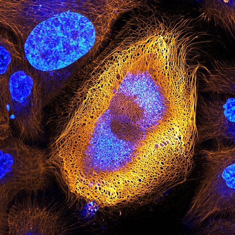
Immortalized human skin cells (HaCaT keratinocytes) expressing fluorescently tagged keratin by Dr. Bram van den Broek, Andriy Volkov, Dr. Kees Jalink, Dr. Reinhard Windoffer & Dr. Nicole Schwarz. Confocal, 40x magnification. 1st place.
Nikon recently unveiled the winners of its annual photomicrography contest, which celebrates the hidden world only seen under a light microscope. For 43 years, scientists, doctors, and enthusiasts have entered their best work into the Nikon Small World Photomicrography Competition, bringing a new worldview to the public.
With over 2,000 entries from 88 countries, the 2017 competition shows a wide array of subjects and techniques. But in the end, it was Dr. Bram van den Broek of The Netherlands Cancer Institute who took home top prize for his image of a skin cell expressing an excessive amount of keratin. He came across the cell while conducting research on the dynamics of keratin filaments.
“There are more than 50 different keratin proteins known in humans. The expression patterns of keratin are often abnormal in skin tumor cells, and it is thus widely used as a tumor marker in cancer diagnostics,” said Dr. van den Broek. “By studying the ways different proteins like keratin dynamically change within a cell, we can better understand the progression of cancers and other diseases.”
Other dynamic images include mold growing on a tomato, a close-up view of a daddy longlegs' eye, and the colorful fractured plastic of a credit card. Stunning in their clarity and artistic in their abstraction, the competition proves just how magical the hidden world can be. “What I most enjoy about this competition is that a larger audience can appreciate the beautiful complexity and diversity of the world unseen by the naked eye,” said van den Broek.
A Small World Exhibit with highlights from the contest travels to museums and science centers throughout the United States. Check here for the schedule.
The winners of the 2017 Nikon Small World Photomicrography Competition showcase the best of life as seen through the light microscope.
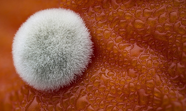
Mold on a tomato by Dean Lerman. Reflected Light and Focus Stacking, 3.9x magnification.
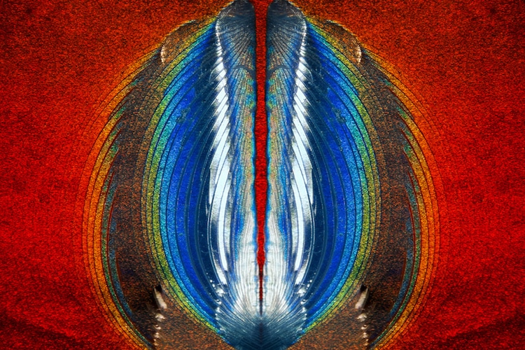
Plastic fracturing on credit card hologram by Steven Simon. Oblique Illumination, 10x magnification. 11th place.
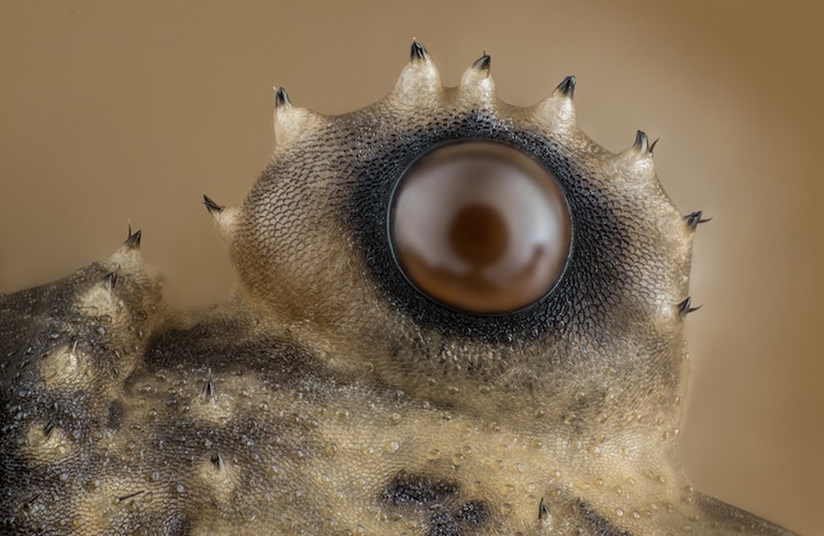
Daddy longlegs eye by Charles B. Krebs. Reflected Light and Image Stacking, 20x magnification.
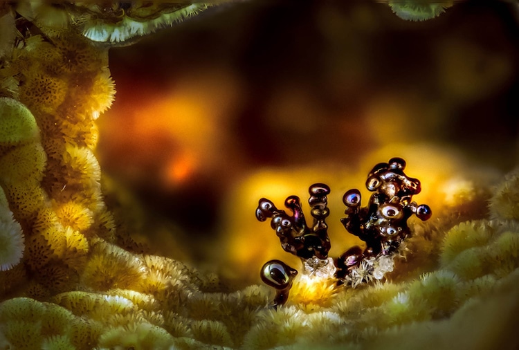
Nsutite and Cacoxenite (minerals) by Emilio Carabajal Márquez. Image stacking,
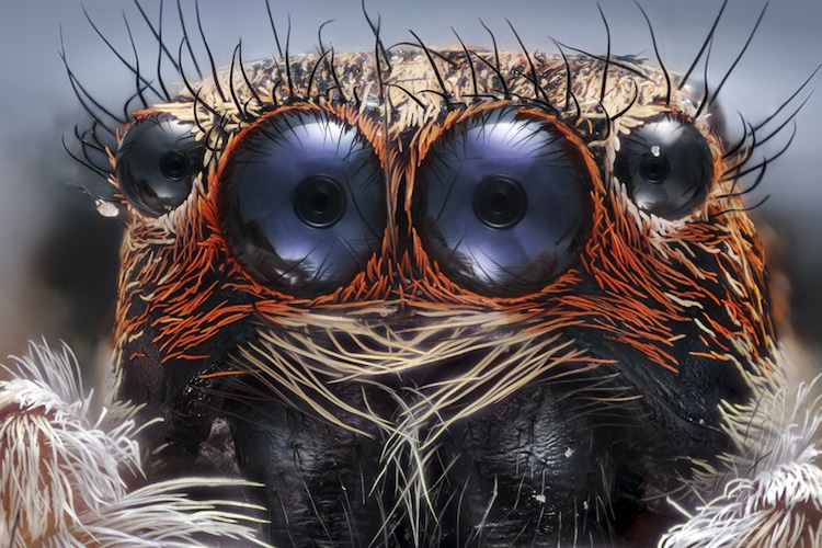
Jumping spider by Emre Can Alagöz. Reflected light, 6x magnification.
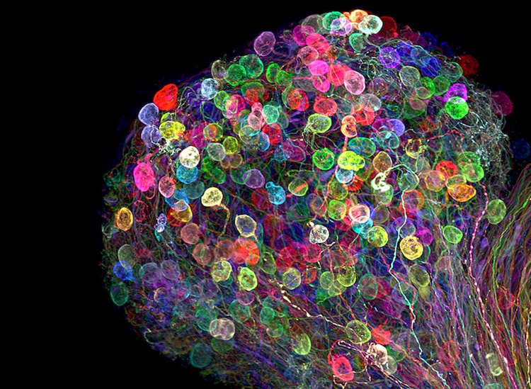
Individually labeled axons in an embryonic chick ciliary ganglion by Dr. Ryo Egawa. Confocal,Tissue Clearing, Brainbow (labeling technique), 30x magnification. 7th place.
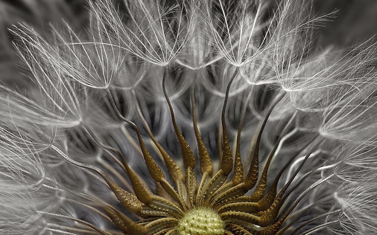
‘Senecio vulgaris' (a flowering plant) seed head by Dr. Havi Sarfaty. 2x magnification. 2nd place.
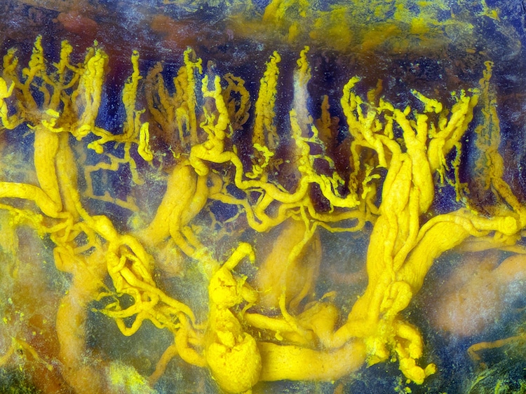
Human tongue blood vessels injected with lead chromate by Frank Reiser. Reflected Light and Focus Stacking, 100x magnification.

Growing cartilage-like tissue in the lab using bone stem cells (collagen fibers in green and fat deposits in red) by Catarina Moura, Dr. Sumeet Mahajan, Dr. Richard Oreffo & Dr. Rahul Tare. Second Harmonic Generation (SHG), Coherent Anti-Stokes Raman Scattering (CARS), 20x magnification for collagen; 40x magnification for fat deposits. 9th place.
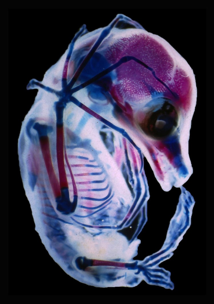
3rd trimester fetus of fruit bat by Dr. Rick Adams. Darkfield and Stereomicroscopy, 18x magnification. 15th place.
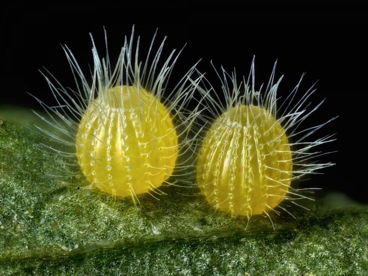
Common Mestra butterfly eggs, laid on a leaf of a Noseburn plant by David Millard. Incident Illumination and Image Stacking, 7.5x magnification. 14th place.
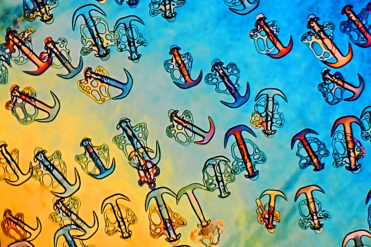
Sea-cucumber skin by Christian Gautier. Polarized Light, 100x magnification. 18th place.
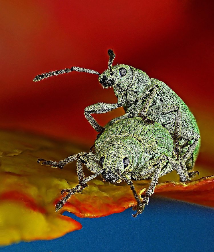
Weevil by Dr. Csaba Pintér. Stereomicroscopy, 80x magnification.
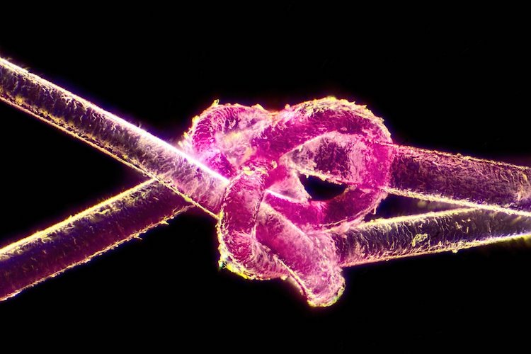
Dyed human hair by Harald K. Andersen. Darkfield, 40x magnification. 17th place.
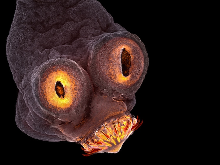
Tapeworm everted scolex by Teresa Zgoda. Confocal, 200x magnification. 4th place.


