The winners and honourably-mentioned images were recently announced for Nikon's 2015 Small World Photomicrography Competition. Submissions were collected from professionals and amateurs alike; the contest is open to anyone with a passion for microscopy and photography. With a rich pool of applicants from 83 different countries submitting over 2,000 entries, the competition was incredibly tough and the calibre of photography that accumulated certainly reflects this. Winners were selected based on artistic quality as well as exceptional scientific technique.
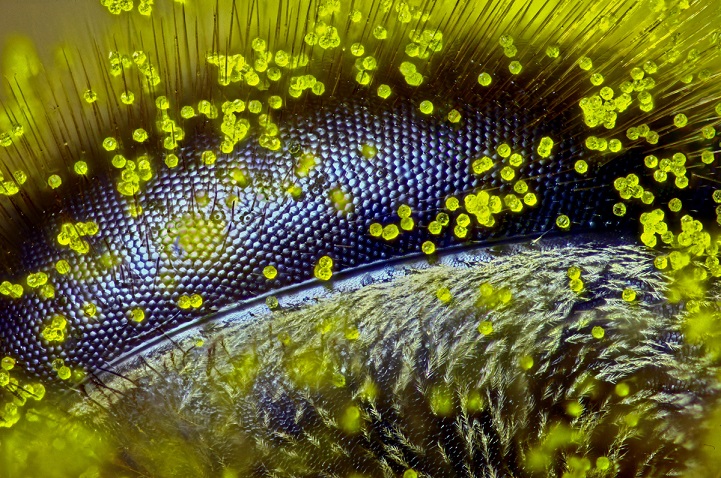
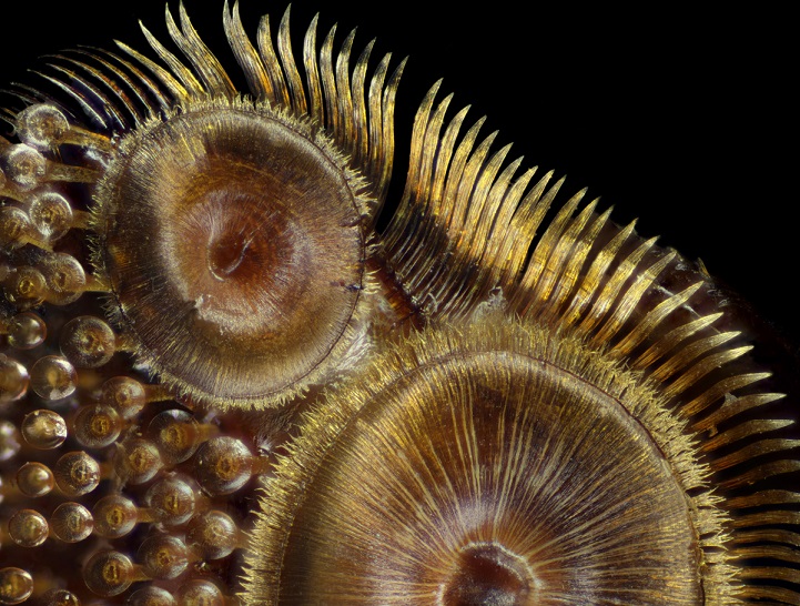
The winning image, seen above, was submitted by Raph Grimm from Queensland, Australia, and gives us a glimpse into the world as seen through the eye of a honeybee. The image took over 4 hours to capture, and the visually striking photograph is a testament to Grimm's painstaking technical labours. While bee populations continue to decline worldwide, Grimm hopes his image can serve as a voice for the tiny pollinators that are so vital to global agriculture, and inspire us to continue to respect and protect our planet.
Regarded as one of the leading platforms for “showcasing the beauty and complexity of life through the lens of a light microscope,” Nikon's Small World offers a rarely seen perspective of the microscopic world. For more incredible photos, you can check out some of the previous year's winning entries online.
Above: Winning Image- The eye of a honeybee (Apis mellifera) covered in dandelion pollen (120x) by Ralph Claus Grimm, Queensland, Australia
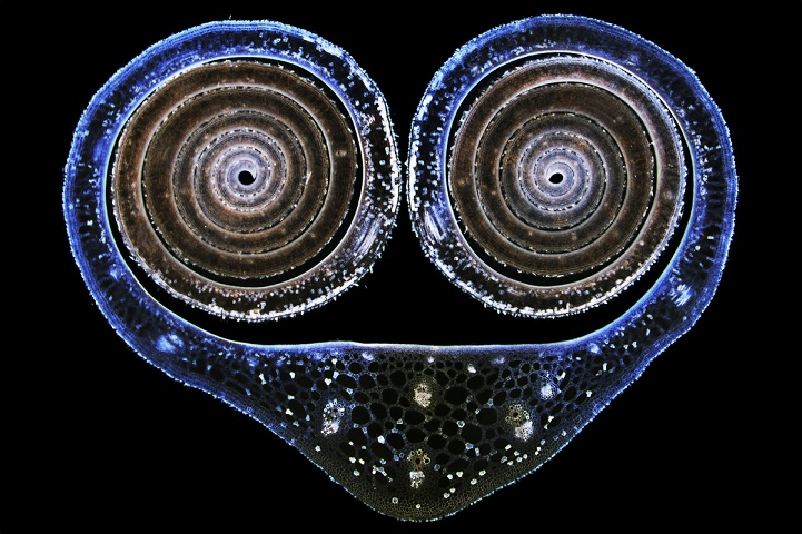
Image of Distinction- Cross section of a water lily leaf bud (Nupha lutea) (12.5x) by Dr. David Maitland, Feltwell, United Kingdom
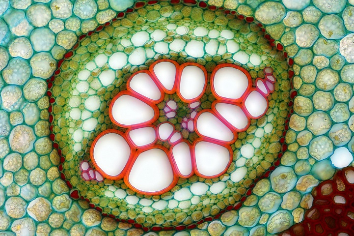
Image of Distinction- Transverse section of an ostrich fern (250x) by Anatoly Mikhaltsov, Omsk, Russia
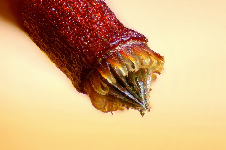
6th Place- Spore capsule of a moss (Bryum sp.) by Henri Koskinen, Helsinki, Finland

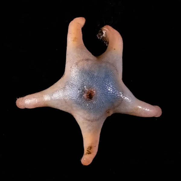
12th Place- Developing sea mullet (Mugil cephalus) embryos (40x) by Hannah Sheppard-Brennand, New South Wales, Australia
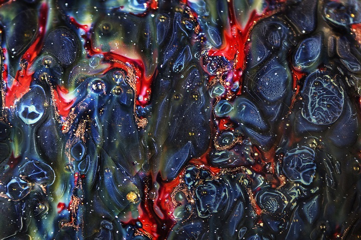
Image of Distinction- Detail of ancient Chinese pottery from the Song Dynasty (960-1126 AD) (4x) by Yvonne (Yi-Chieh) Lu, New York, USA

Image of Distinction- The feeding structure (radula) of a limpet, a kind of aquatic snail (40x) by Michael Crutchley, Wales, United Kingdom
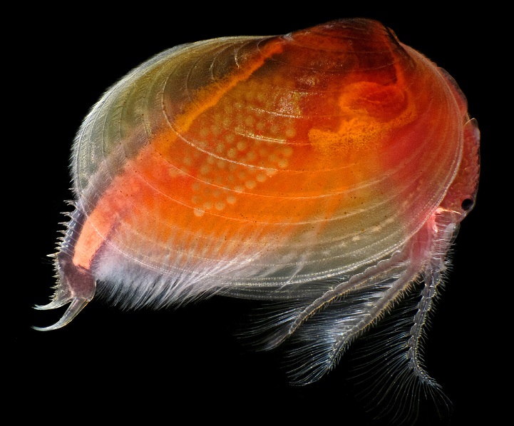
10th Place- Clam shrimp (Cyzicus mexicanus), live specimen (25x) by Ian Gardiner, Alberta, Canada
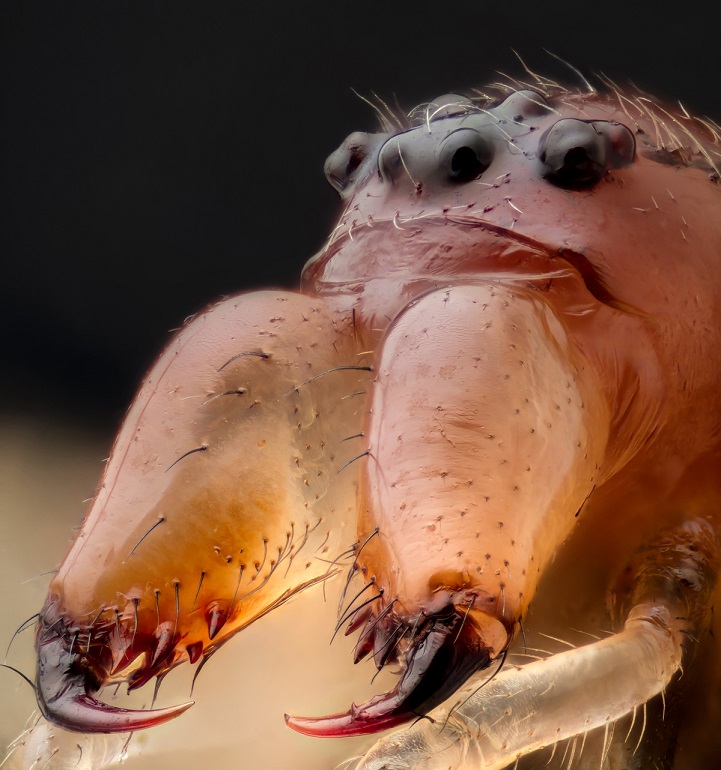
Image of Distinction- Jaws and head of a long-jawed spider (Metellina sp.) (10x) by Geir Drange, Asker, Norway
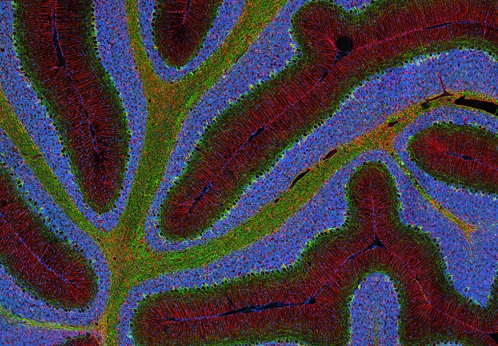
Image of Distinction- Triple-labeled rat cerebellum (100x) by Thomas Deerinck, California, USA
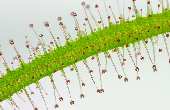
13th Place- Tentacles of a carnivorous plant (Drosera sp.) (20x) by Jose Almodovar, Puerto Rico, USA
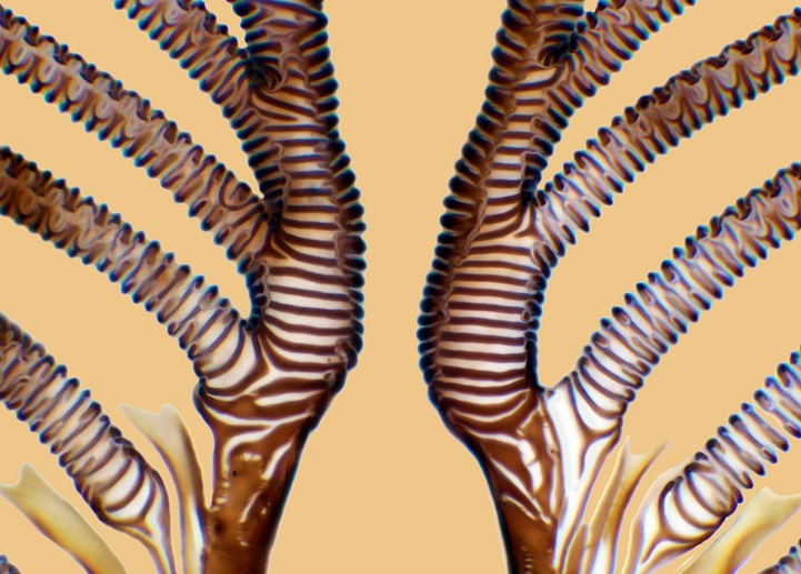
Image of Distinction- Mouth parts (pseudo trachea) of a blowfly (Calliphora vomitoria) (750x) by Raymond Morrison Sloss, Oxfordshire, United Kingdom
20th Place- Suction cups on a diving beetle (Dytiscus sp.) foreleg (50x) by Frank Reiser, New York, USA
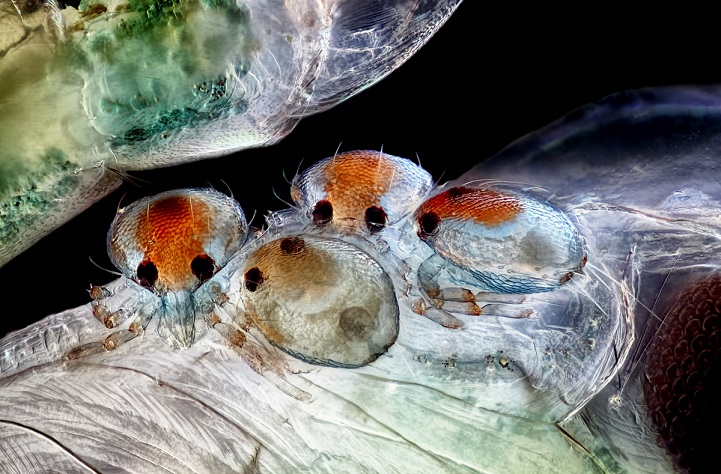
Honorable Mention- Mites on insect pupa (20x) by Rogelio Moreno Gill, Panama
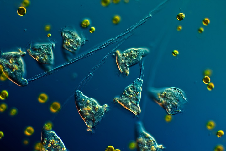
Image of Distinction- Colony of single-celled organisms (Carchesium ciliates) (160x) by Arturo Agostino, Reggio Calabria, Italy
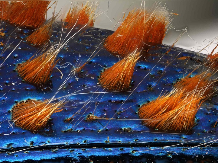
Honorable Mention- Detail of jewel beetle (Coleoptera Buprestidae) (32x) by Dr. Luca Toledano, Verona, Italy
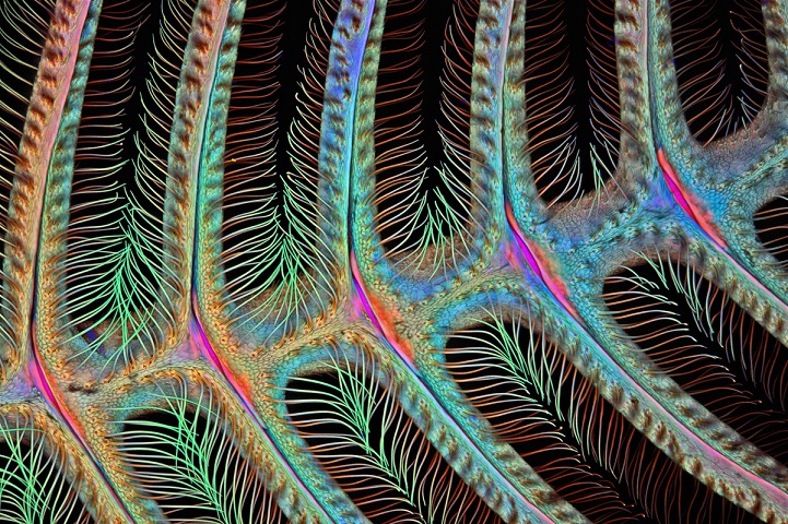
Image of Distinction- Antenna of a male moth (Anisota sp.) (100x) by Dr. Igor Siwanowicz, Virginia, USA