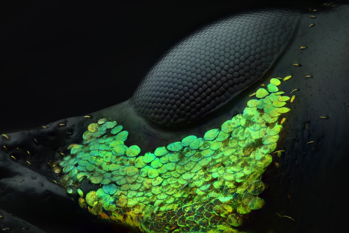
Eye of a “Metapocyrtus subquadrulifer” beetle by Yousef Al Habshi (Abu Dhabi, United Arab Emirates). Reflected Light, 20x (objective lens magnification). 1st place.
For the 44th year, Nikon celebrates the invisible world with its Small World Photomicrography Competition. As imaging and microscope technologies evolve, scientists, professional photographers, and hobbyists continue to push the boundaries of micrography. For the 2018 competition, nearly 2,500 scientists and artists from 89 countries submitted their work.
After being evaluated on originality, informational content, technical proficiency, and visual impact, Yousef Al Habshi from Abu Dhabi was declared the winner. His incredible image of an Asian Red Palm weevil's eye is a close up look at the insect's striking anatomy. This tiny beetle, which is found in the Philippines, typically measures less than 11 mm (0.43 in). Iridescent green disks, which look like sequins, are laid out in a perfectly overlapping pattern across the black body. Al Habshi's final image comes from stacking over 128 micrographs as not to overexpose the skin and scales while still showing off the black body.
“Because of the variety of coloring and the lines that display in the eyes of insects, I feel like I'm photographing a collection of jewelry,” said Al Habshi. “Not all people appreciate small species, particularly insects. Through photomicrography we can find a whole new, beautiful world which hasn't been seen before. It's like discovering what lies under the Ocean's surface.”
The entire top twenty makes use of a wide variety of techniques to bring their photomicrographs to life. Familiar names like Justin Zoll and Can Tunçer are included in the list for their images of crystallized amino acids and peacock feathers, respectively. Whether exploring a human teardrop or seeing how a tiny plant-sucking insect protects itself in a foam bubble, the winning images open up a new window to our world.
The winners of the Nikon Small World Photomicrography Competition will have their work shown during a traveling exhibition around the United States and Canada. They will also be included in a full-color calendar.
For the past forty-four years, Nikon Small World has celebrated the best photomicrography across the globe.
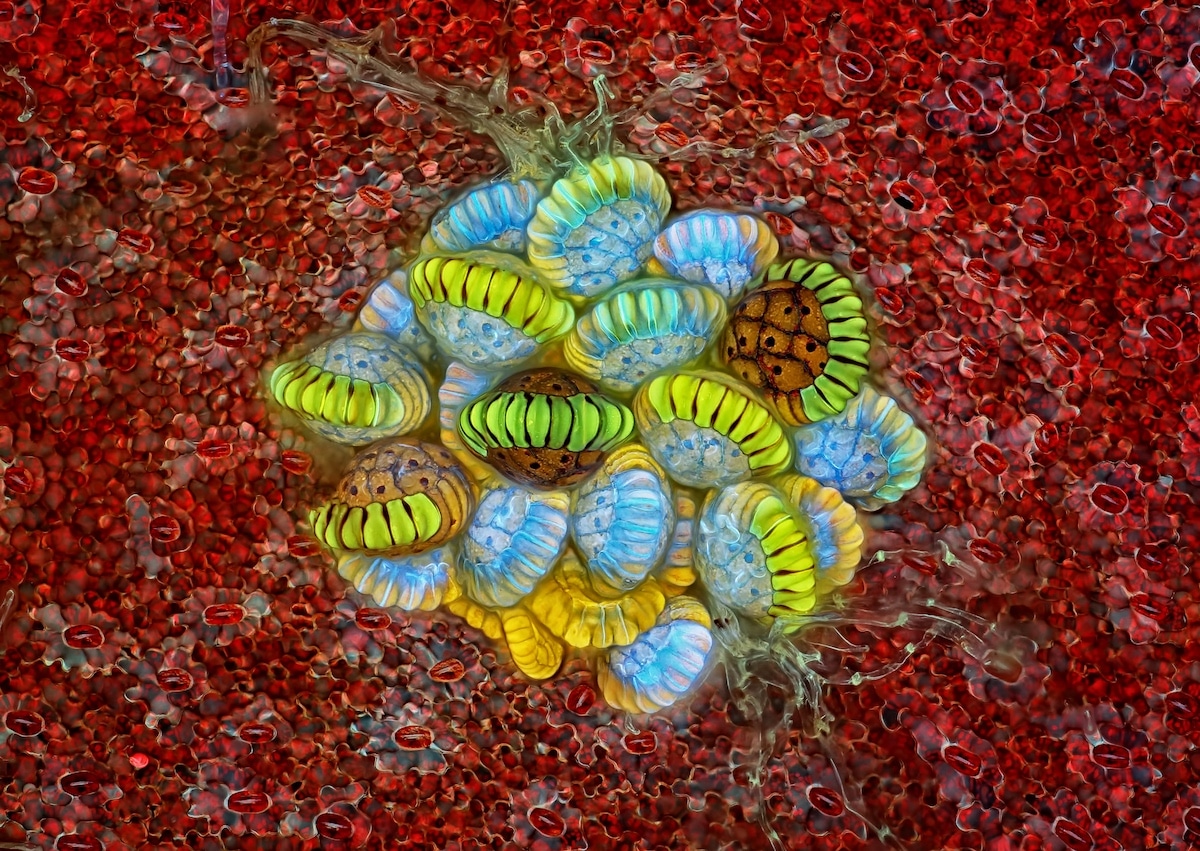
“Fern sorus” (structures producing and containing spores) by Rogelio Moreno (Panama, Panama). Autofluorescence 10x (objective lens magnification). 2nd place.
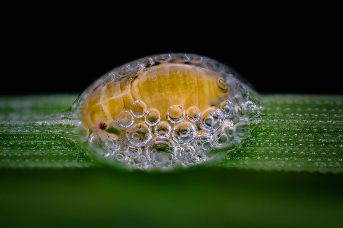
Spittlebug nymph in its bubble house by Saulius Gugis (Naperville, Illinois). Focus Stacking, 5x (objective lens magnification). 3rd place.
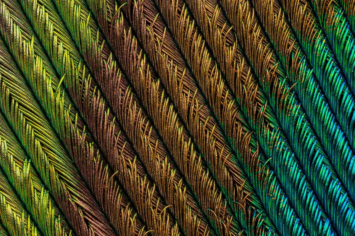
Peacock feather section by Can Tunçer (İzmir, Turkey). Focus Stacking, 5x (objective lens magnification). 4th place.
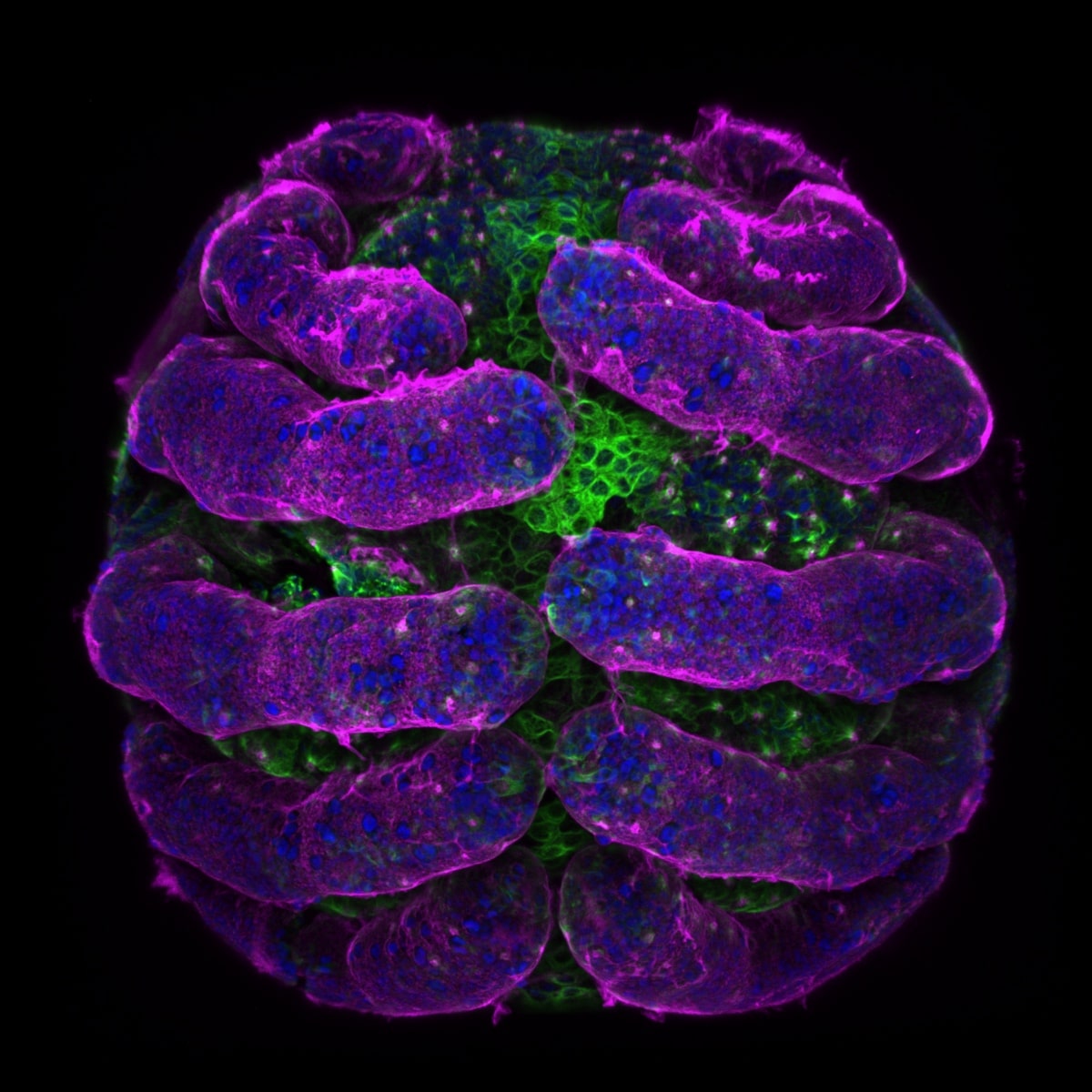
“Parasteatoda tepidariorum” (spider embryo) stained for embryo surface (pink), nuclei (blue) and microtubules (green) by Dr. Tessa Montague (Harvard University, Department of Molecular and Cellular Biology, Cambridge, Massachusetts). Confocal, 20x (objective lens magnification). 5th place.
Over 2,500 scientists and artists from 89 countries entered the 2018 photo competition.
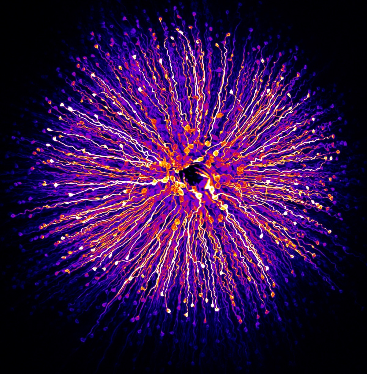
Primate foveola (central region of the retina) by Hanen Khabou (Vision Institute, Department of Therapeutics, Paris, France). Fluorescence, 40x (objective lens magnification). 6th place
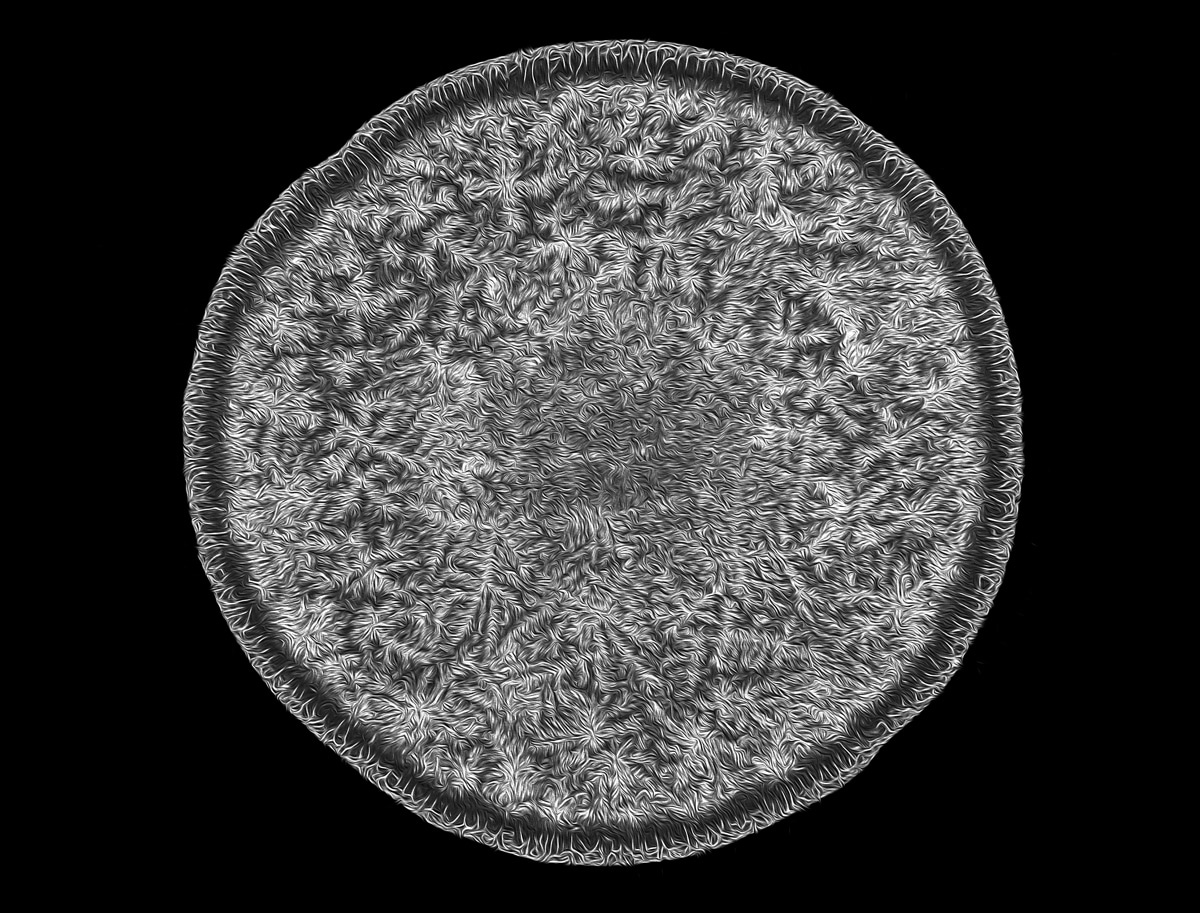
Human tear drop by Norm Barker (Johns Hopkins School of Medicine, Department of Pathology & Art as Applied to Medicine, Baltimore, Maryland). Darkfield , 5x (objective lens magnification). 7th place.
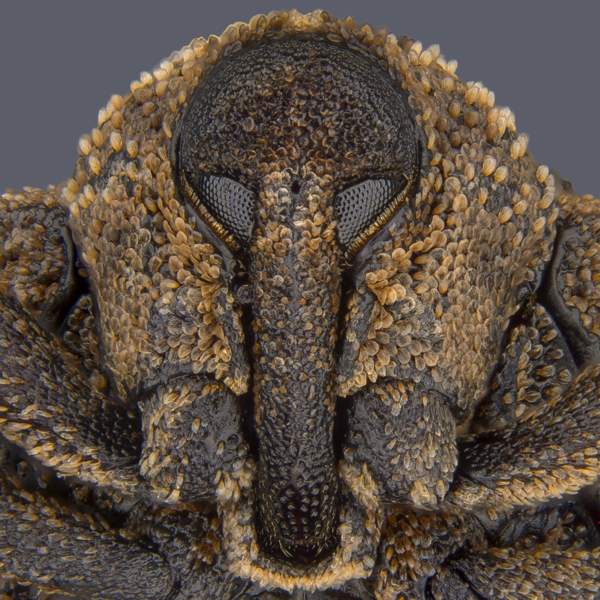
Portrait of “Sternochetus mangiferae” (mango seed weevil) by Pia Scanlon (Government of Western Australia, Department of Primary Industries and Regional Development South Perth, Western Australia). Stereomicroscopy, Image Stacking, 1x (objective lens magnification). 8th place.
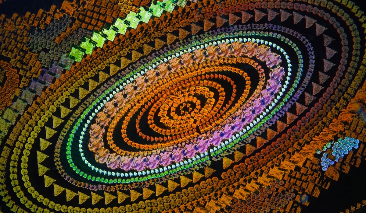
Security hologram by Dr. Haris Antonopoulos (Athens, Greece). Darkfield Epi-illumination, 10x (objective lens magnification). 9th place.
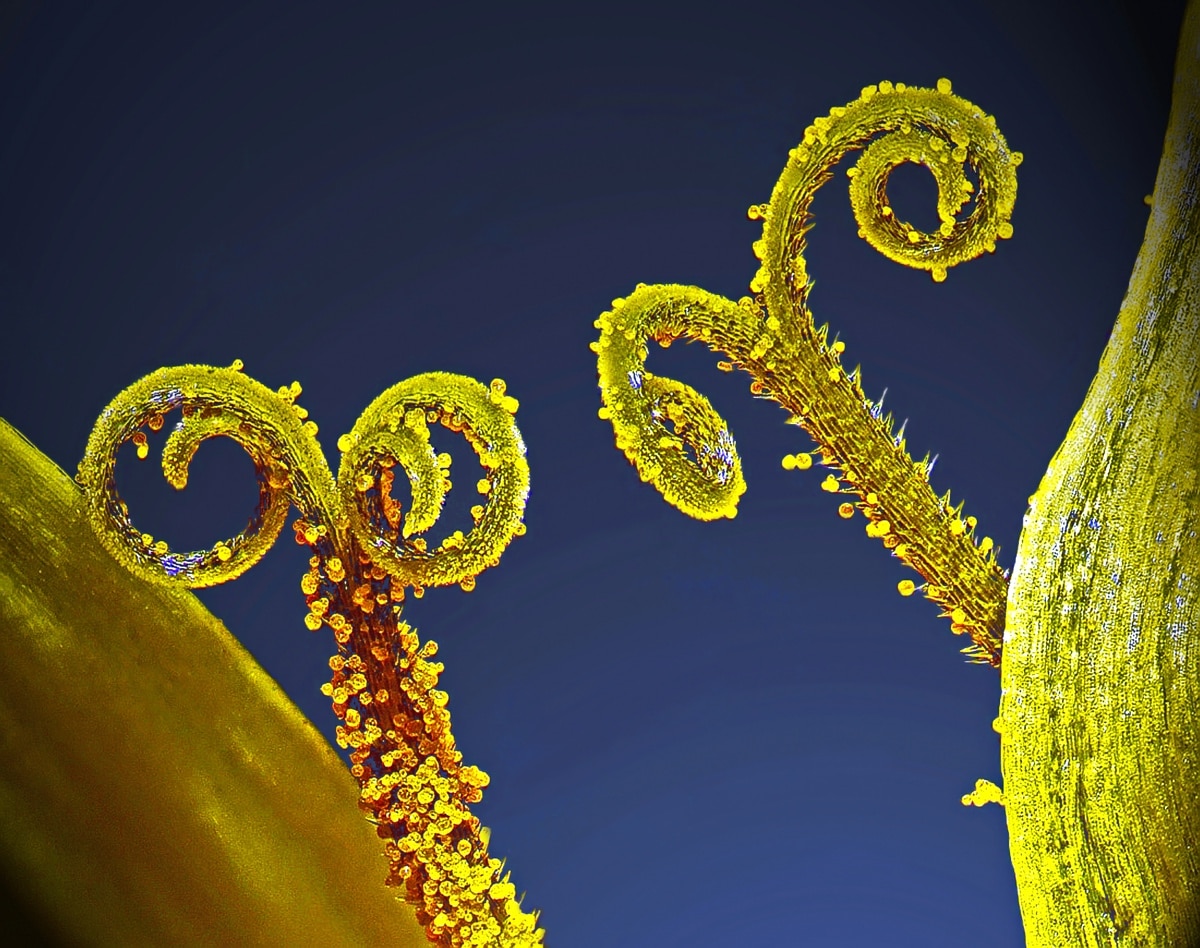
Stalks with pollen grains by Dr. Csaba Pintér (University of Pannonia, Georgikon Faculty, Department of Plant Protection, Keszthely, Hungary). Focus Stacking, 3x (objective lens magnification). 10th place.
“The Nikon Small World competition is now in its 44th year, and every year we continue to be astounded by the winning images,” said Eric Flem, Communications Manager, Nikon Instruments.
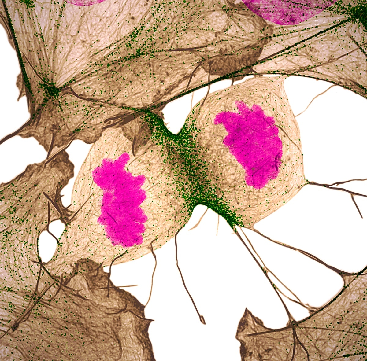
Human fibroblast undergoing cell division, showing actin (gray), myosin II (green) and DNA (magenta) by Nilay Taneja & Dr. Dylan Burnette (Vanderbilt University, Department of Cell and Developmental Biology, Nashville, Tennessee). Structured Illumination Microscopy, 60x (objective lens magnification). 11th place.
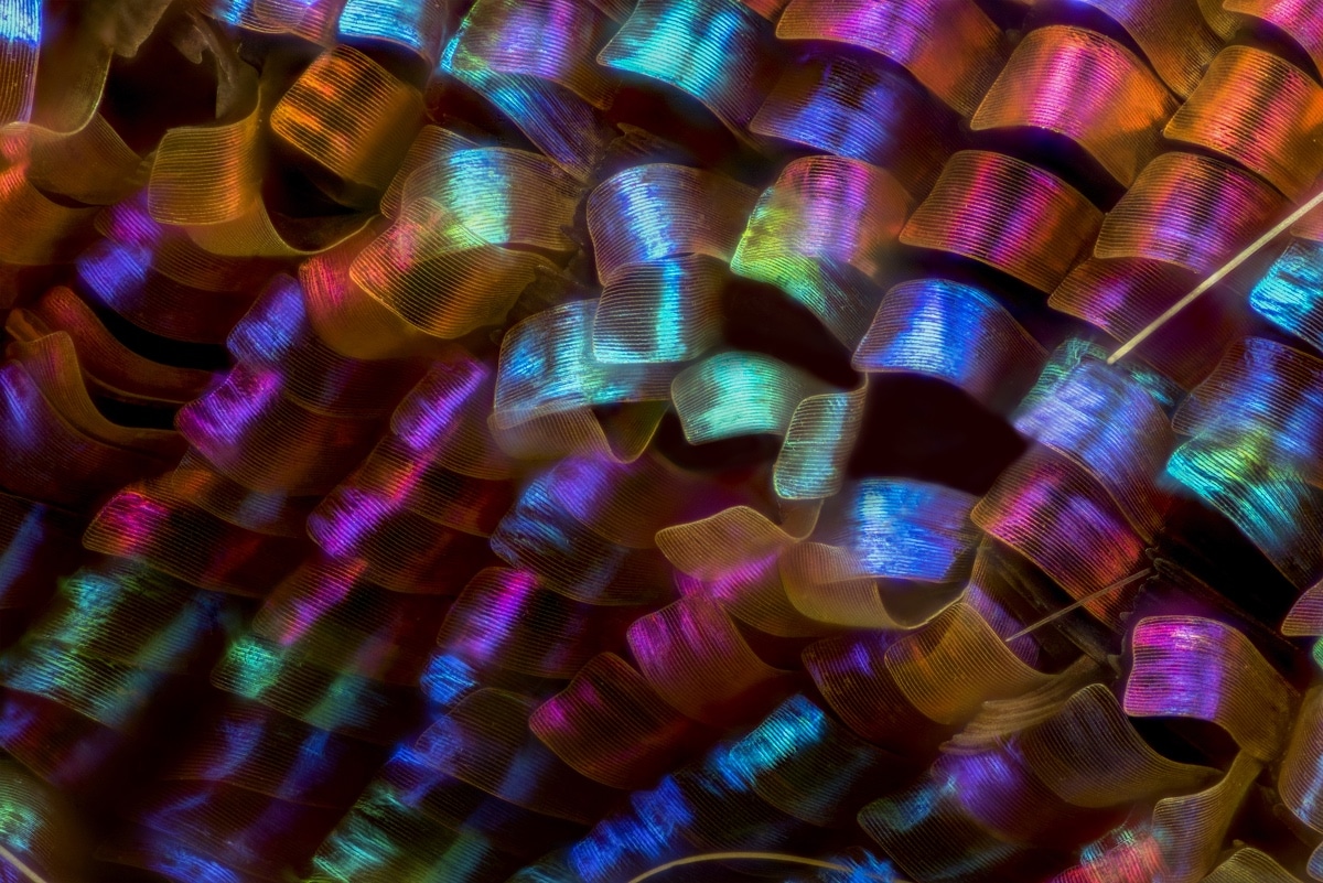
“Urania ripheus” (butterfly) wing scales by Luciano Andres Richino (Punto NEF Photography, Ramos Mejia, Buenos Aires Province, Argentina). Image Stacking, 20x (objective lens magnification). 12th place.
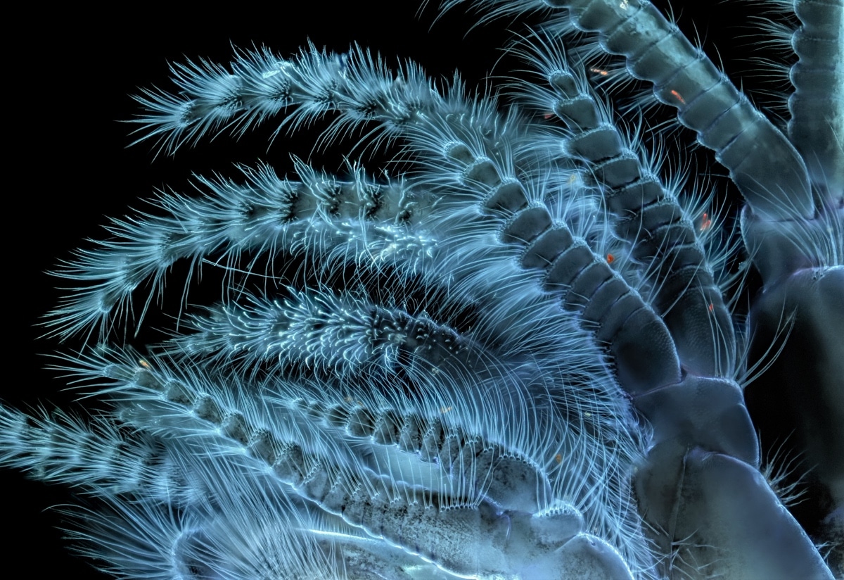
“Balanus glandula” (acorn barnacle) by Charles Krebs (Charles Krebs Photography, Issaquah, Washington). Autofluorescence, 5x (objective lens magnification). 13th place.
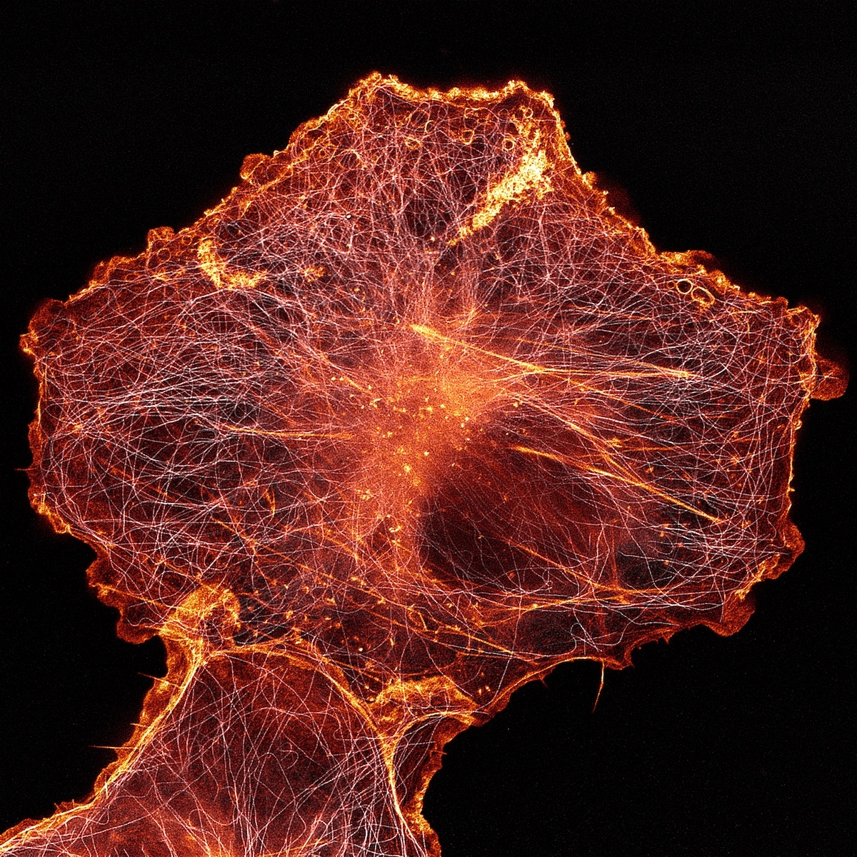
African green monkey cell (COS-7) stained for actin and microtubules by Andrew Moore & Dr. Erika Holzbaur (University of Pennsylvania, Department of Physiology, Philadelphia, Pennsylvania). Stimulated Emission Depletion (STED) Microscopy, 100x (objective lens magnification). 14th place.
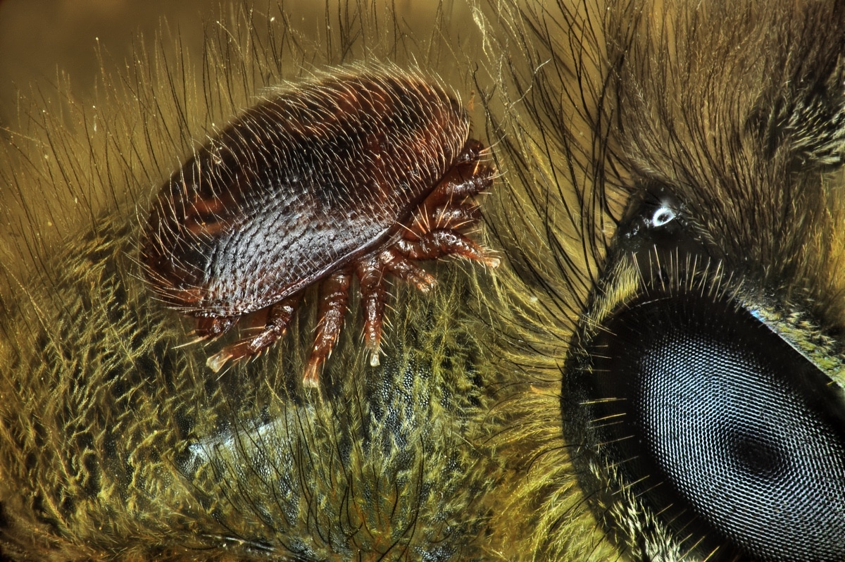
“Varroa destructor” (mite) on the back of “Apis mellifera” (honeybee) by Antoine Franck (CIRAD – Agricultural Research for Development, Saint Pierre, Réunion, Reunion Island). Focus Stacking, 1x (objective lens magnification). 15th place.
“Imaging and microscope technologies continue to develop and evolve to allow artists and scientists to capture scientific moments with remarkable clarity.”
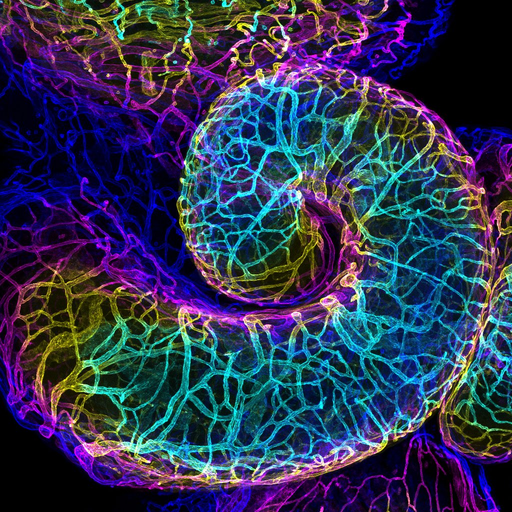
Mouse oviduct vasculature by Dr. Amanda D. Phillips Yzaguirre (Salk Institute for Biological Studies, La Jolla, California). Confocal, 10x (objective lens magnification). 16th place.
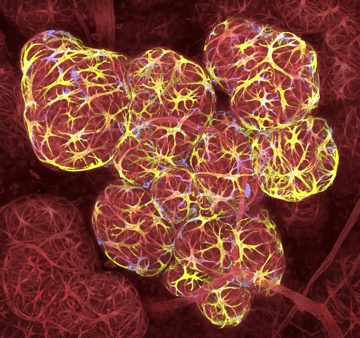
Breast tissue in lactation: Milk filled spheres (red) surrounded by tiny muscle cells that squeeze out milk (yellow) and immune cells that monitor for infection (blue) by Caleb Dawson (The Walter and Eliza Hall Institute of Medical Research, Department of Stem Cells and Cancer, Melbourne, Australia). 3D Confocal Microscopy, 63x (objective lens magnification). 17th place.
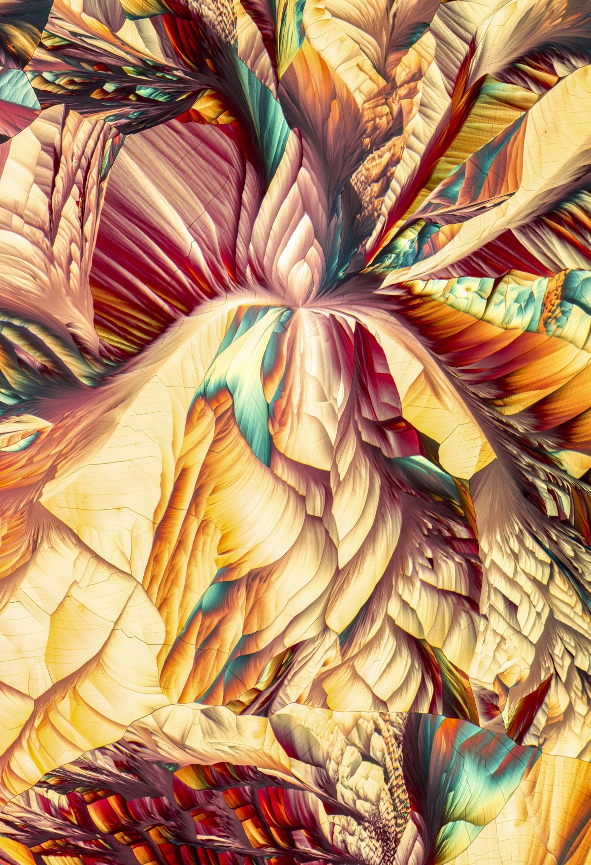
Amino acid crystals (L-glutamine and beta-alanine) by Justin Zoll (Justin Zoll Photography, Ithaca, New York). Polarized Light, Image Tiling, 4x (objective lens magnification). 18th place.
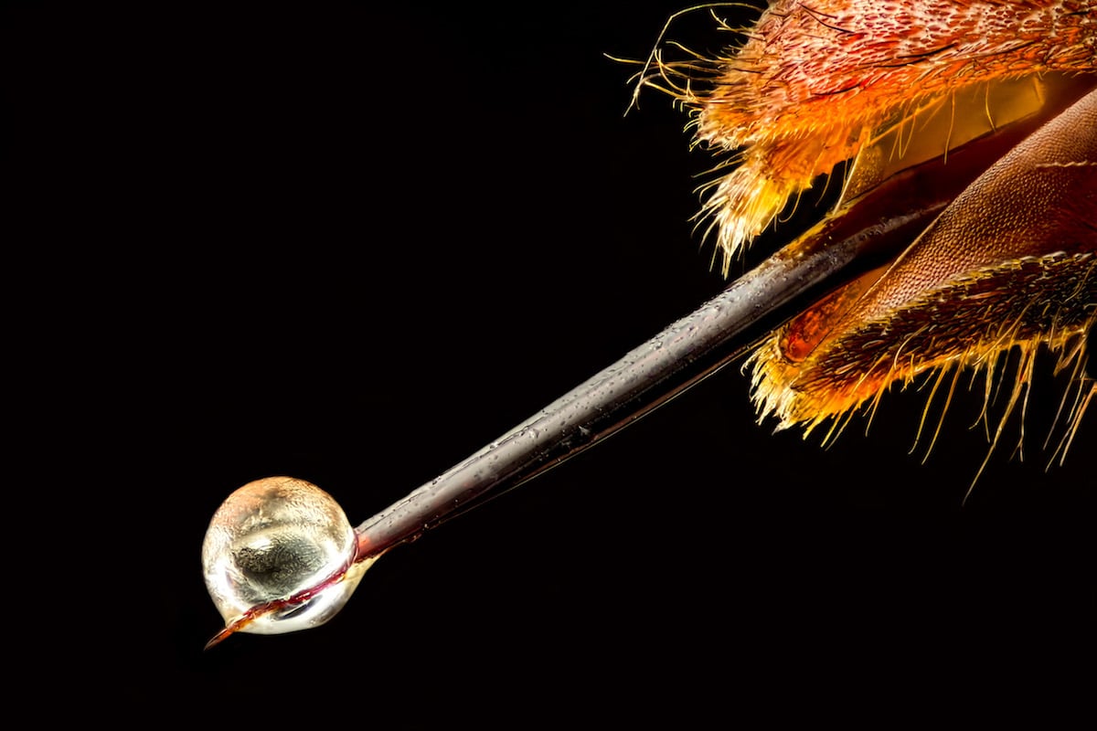
“Vespa velutina” (Asian hornet) with venom on its stinger by Pierre Anquet (La Tour-du-Crieu, Ariège, France). Reflected Light, Focus Stacking, 6.3x (objective lens magnification). 19th place.
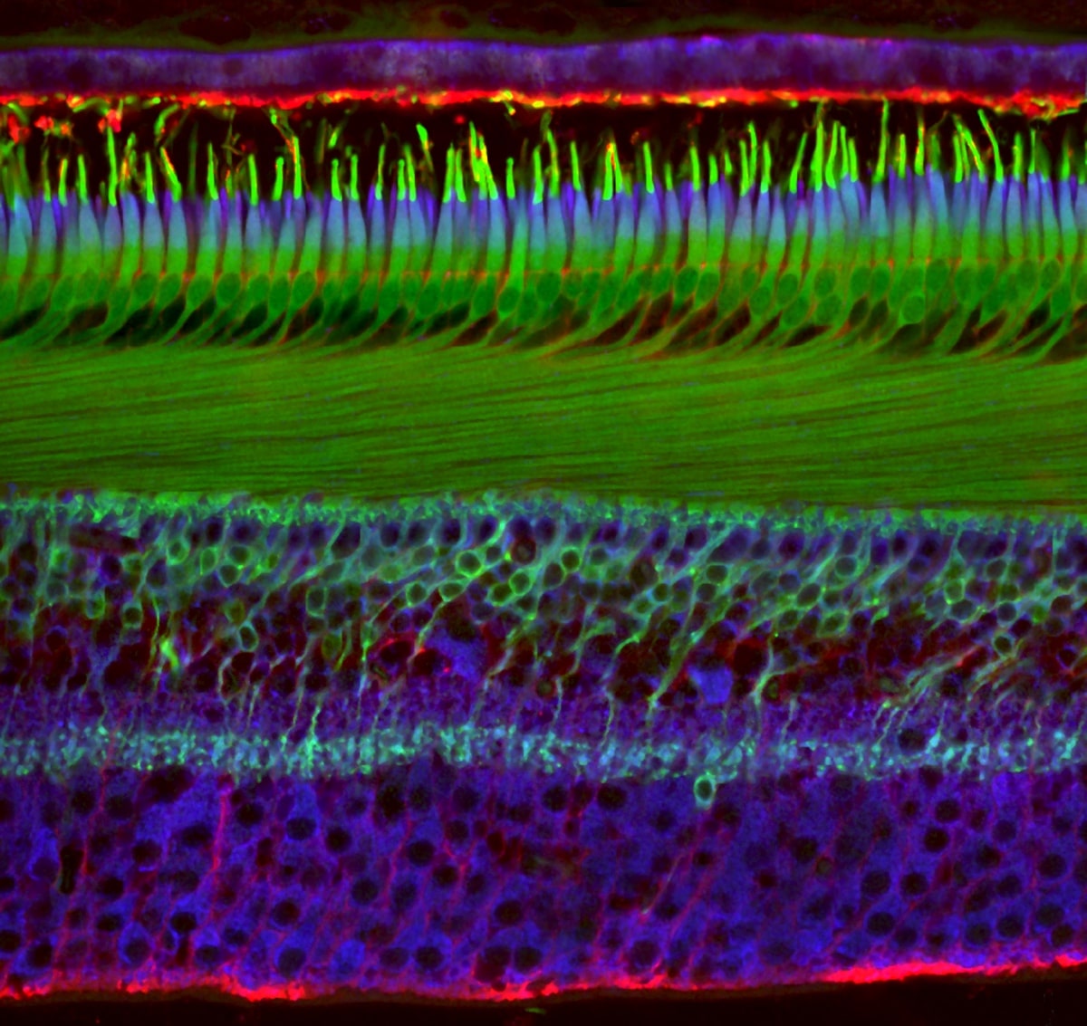
Human retina by Dr. Nicolás Cuenca & Isabel Ortuño-Lizarán (University of Alicante, Department of Physiology, Genetics and Microbiology, San Vicente del Raspeig, Alicante, Spain). Immunocytochemistry and Confocal Microscopy, 40x (objective lens magnification). 20th place.


