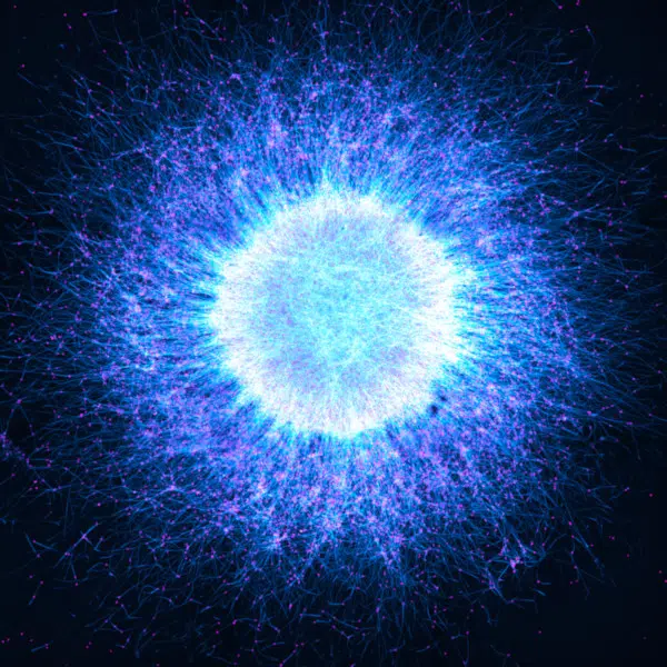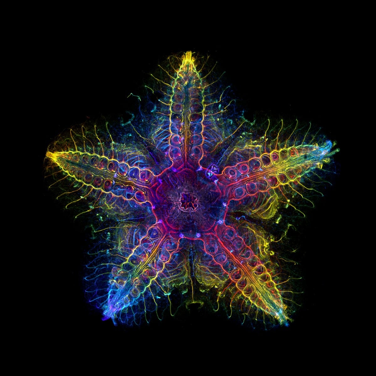
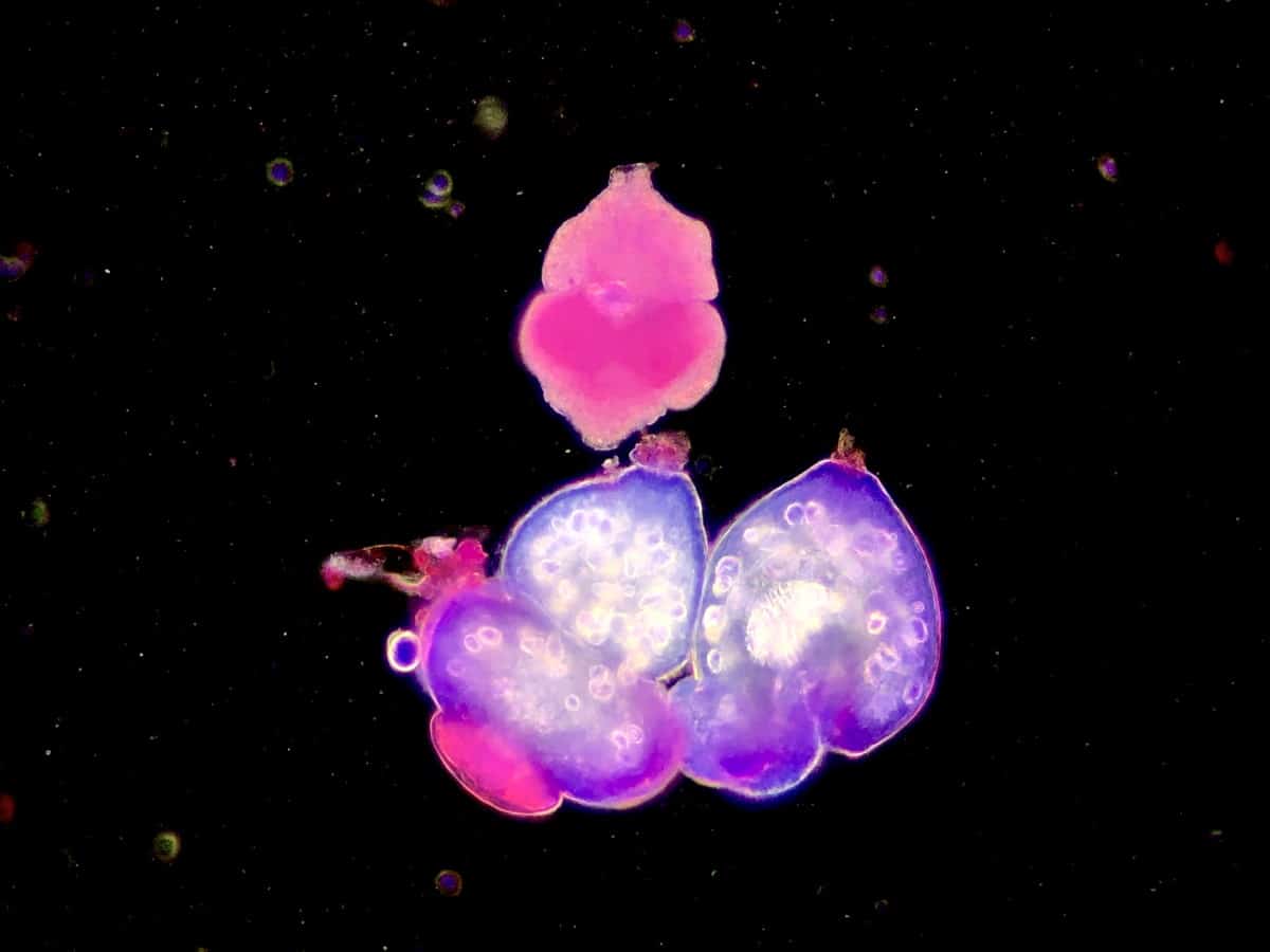
Global Winner, Laurent Formery (USA).
Nervous system of a juvenile sea star (Patiria miniata) about 1 cm wide. Labeled with an antibody against acetylated tubulin after optical clearing, and captured using a color-coded Z-projection.
A look at the inner workings of a sea star won the Global Image of the Year Scientific Light Microscopy Award. The contest, which recognizes the best in scientific imaging worldwide, selected the winner from 640 images submitted by photographers and scientists from 38 countries.
Organized by Evident and Olympus, since 2017, the contest has been an incredible way to highlight the artistry and scientific value of light microscopy. Global winner Laurent Formery was thrilled by the victory, which comes two years after he earned an honorable mention in the contest.
“This is a fantastic feeling,” Formery shared. “Winning the global award feels like an incredible achievement and shows I made progress. I love microscopy and can spend a huge amount of time in front of our confocal microscope, but the very nice samples that I am lucky to work with really make the difference. I work with marine invertebrates, in particular echinoderms (sea stars, sea urchins, and their kind). They are beautiful animals, with a fascinating and aesthetically pleasing fivefold symmetry that is unlike anything else in the animal kingdom. I’m happy that taking images of them helps communicate how much beauty we have in our oceans, and why it is important to know more about them and protect them.”
For the first time, the contest also singled out material sciences and engineering with a special Materials Science prize. Shyam Rathod from India won the inaugural award—and an Olympus SZ61 stereo microscope—for a colorful image showing the crystal of a topical medicine for wart treatment.
Scroll down for more winning images, as well as the honorable mentions from this year's contest and get a glimpse at life under the microscope.
The Global Image of the Year Scientific Light Microscopy Awards highlights the best scientific imaging.
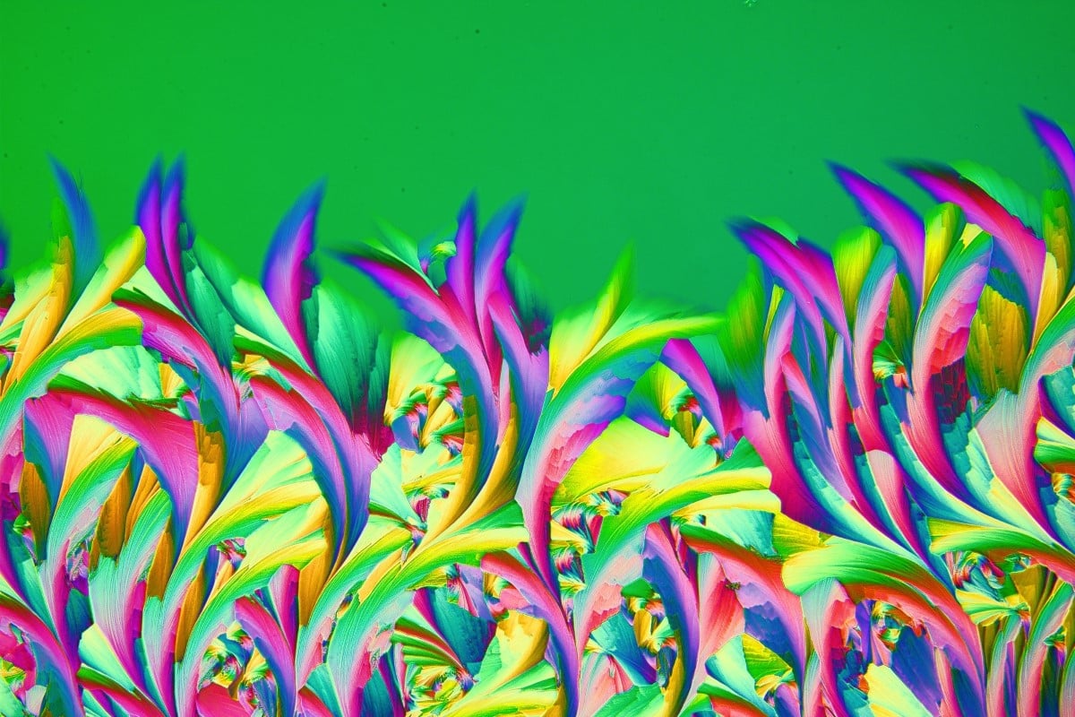
Materials Science Winner, Shyam Rathod (India).
Crystal of a topical medicine for wart treatment named ABE, which is available in Poland. The dew was blown on a microscope slide with a straw and was captured in a single frame. Retarder and a two-cross polarization filter were used to bring out the colors.
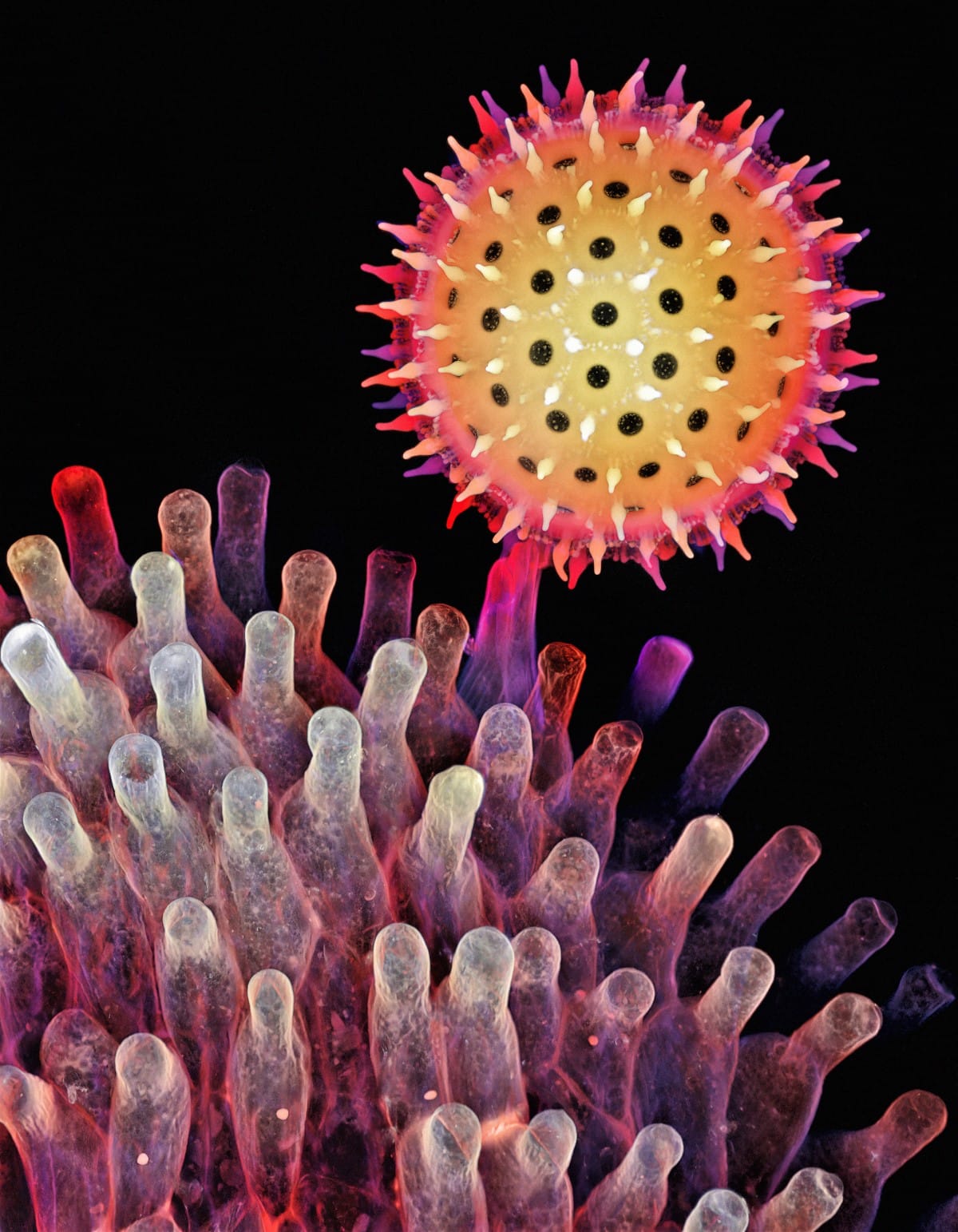
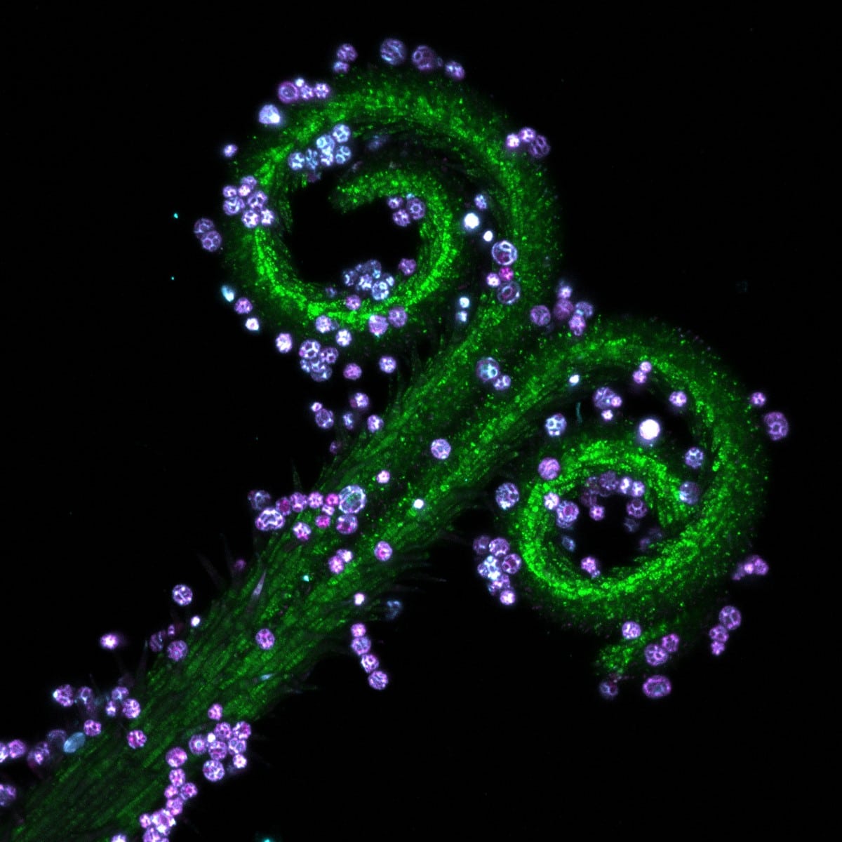
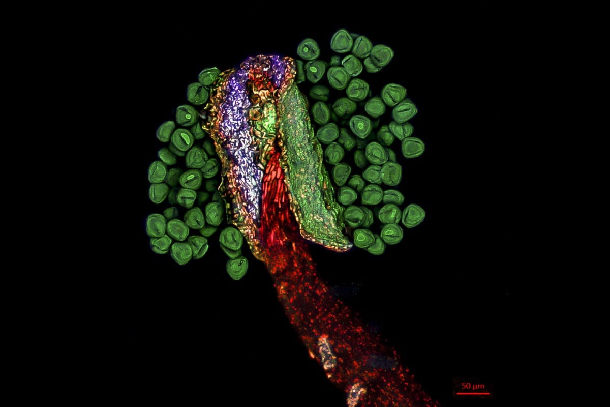
Regional Winners, Americas, Igor Siwanowicz (USA).
Depth color-coded projection showing a germinating pollen grain of a morning glory attached to the stigma.
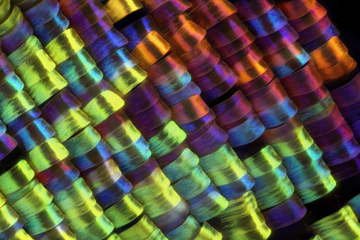
Regional Winner, Europe, the Middle East, and Africa, Javier Ruperez (Spain).
Scales of the wing of the Urania rhipheus moth. Captured at 20X.
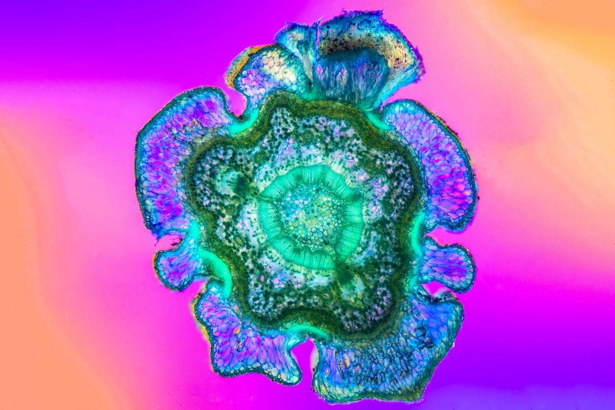
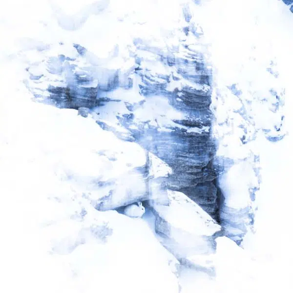
Honorable mention, Robert Berdan (Canada).
Cross section of a blue spruce (Picea pungens) branch. The image is a panorama of several photos that were focus stacked and captured with a 5X objective.
640 images from 38 countries were entered into this year's contest.
Honorable mention, Liu Ruming (China).
Pistils of dandelion were collected and made into slide samples. Different fluorescence patterns of its parts were observed using confocal microscopy.
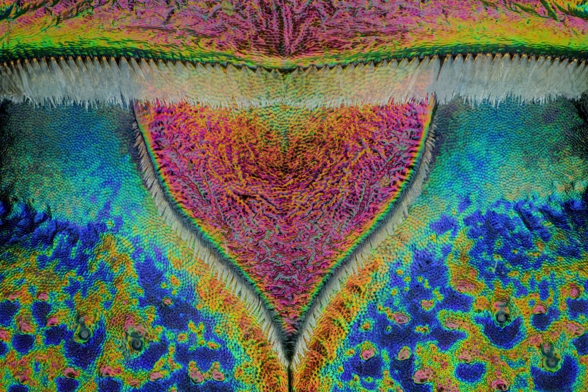
Honorable mention, Thorben Danke (Germany)
Scutellum of a tiger beetle 20:1.
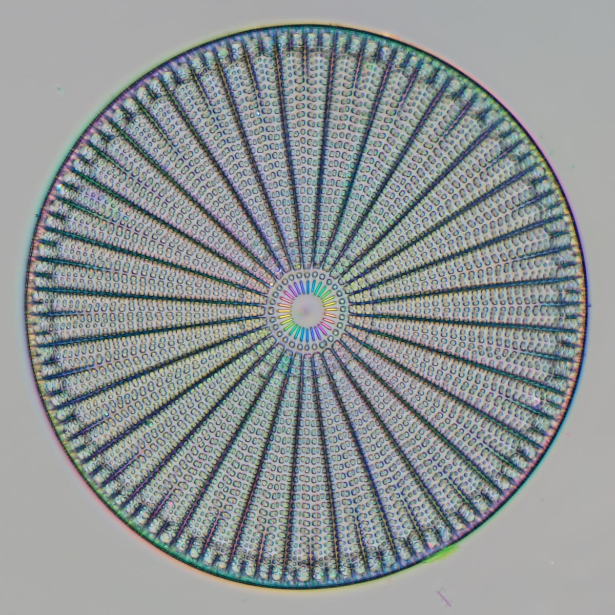
Honorable mention, Michael Shribak (USA)
Diatoms are unicellular organisms, which can be found in the oceans, waterways, and soils of the world.
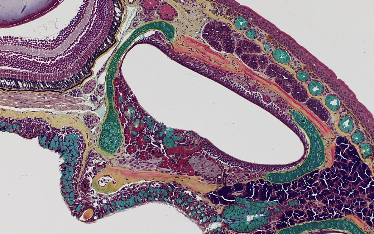
Honorable mention, Katelin Murphy (USA).
Dorsal view of a red-backed salamander skull. Stained with Movat's pentachrome technique.
Regional Winner, Asia-Pacific, Jiao Li (China).
Edelweiss stamens, which were scanned and reconstructed in three dimensions using laser scanning confocal microscopy.
Honorable mention, Ru Jinwei (China).
Fine grained Echinococcus protocephalus, stained with SM and
photographed in dark field.
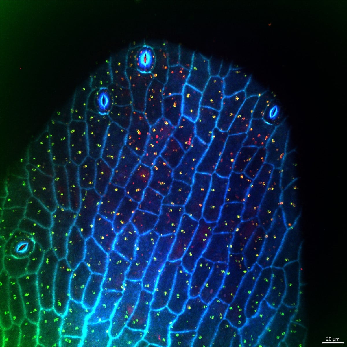
Honorable mention, Mei Yu (China).
Autofluorescence of the tip of the stamens of flowers at 405 nm, 488 nm, and 561 nm, emitting a greenish blue and emerald green color like peacock feathers.
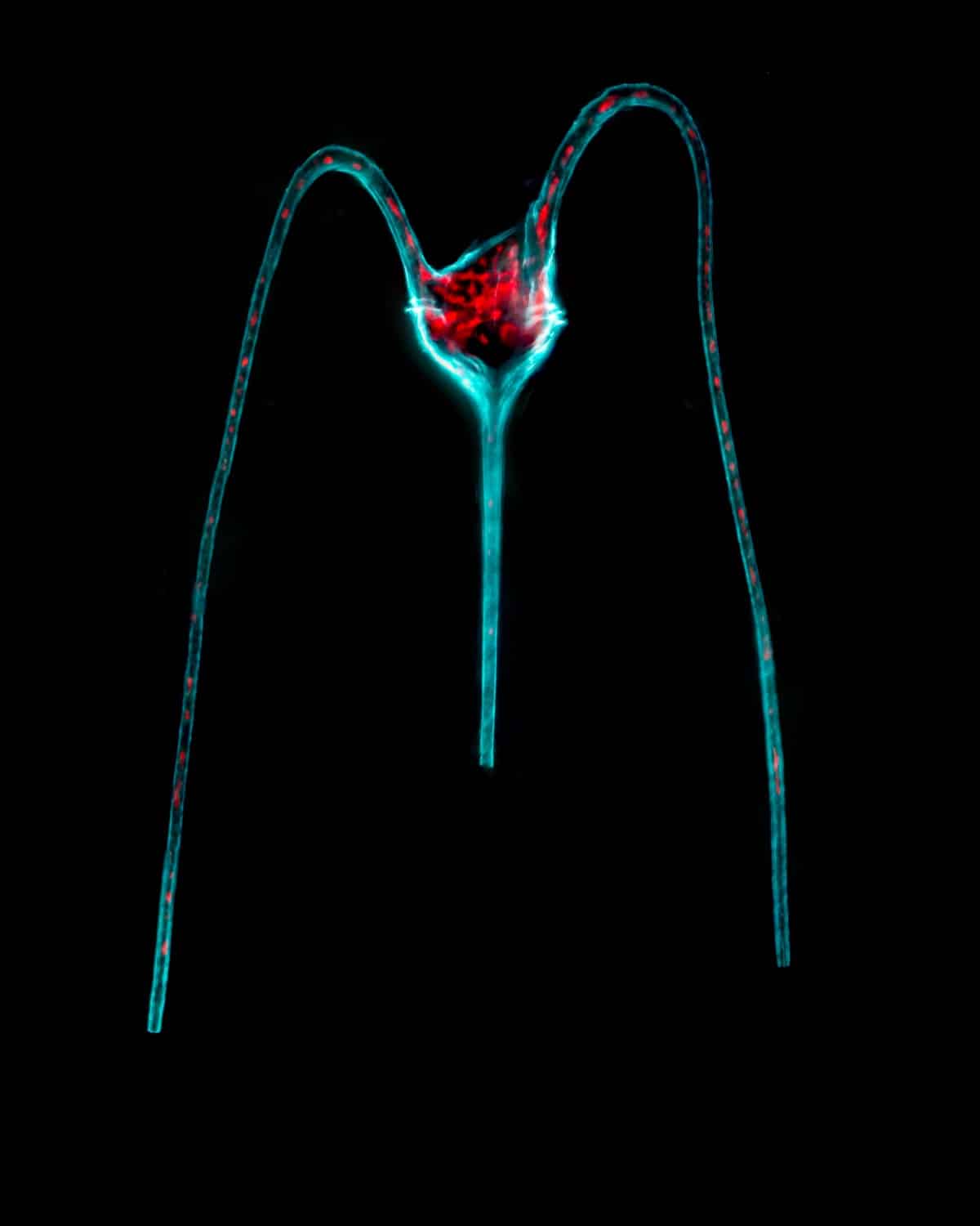
Honorable mention, Uriel Ruiz (Mexico).
Tripos macroceros. A unicellular microalga with three horns. The antapical horns are bent toward the single apical horn. The chlorophyll is displayed in red, where the chloroplasts sit.