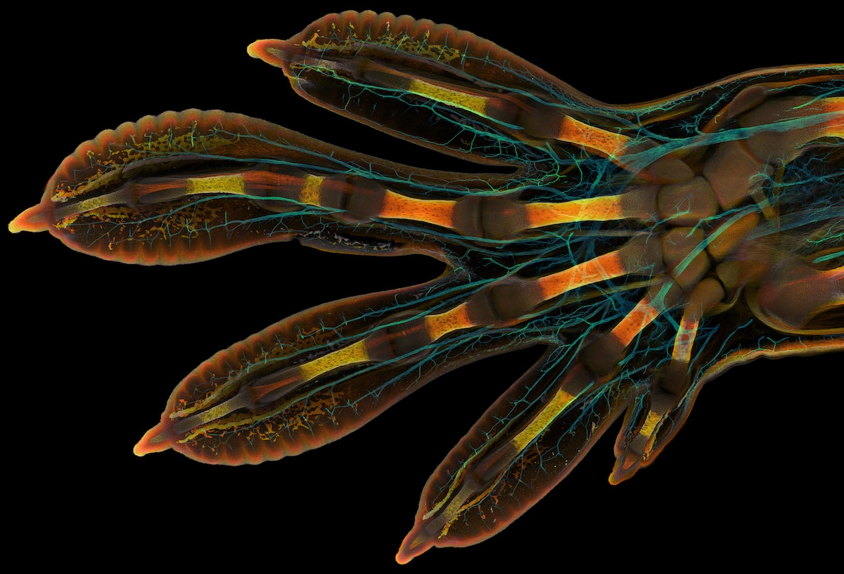
Embryonic hand of a Madagascar giant day gecko (Phelsuma grandis), 1st Place, Grigorii Timin & Dr. Michel Milinkovitch, University of Geneva, Department of Genetics and Evolution, Confocal, 63X (Objective Lens Magnification)
For the past 48 years, the Nikon Small World Photomicrography Competition has been revealing the beauty of a world that can only be seen with a microscope. Every year the contest produces exceptional results and 2022 is no different. Their year’s contest received almost 1,300 entries from 72 countries. The winning image of a Madagascar giant day gecko's embryonic hand is an incredible look at life in development. Taken by Grigorii Timin, with supervision by Dr. Michel Milinkovitch, the photo is the result of painstaking image-stitching.
“This embryonic hand is about 3 mm (0.12 in) in length, which is a huge sample for high-resolution microscopy,” said Timin. “The scan consists of 300 tiles, each containing about 250 optical sections, resulting in more than two days of acquisition and approximately 200 GB of data.”
Thanks to his work, we're able to see the complexity of the gecko. Nerves are stained in a cyan color, while the bones, tendons, ligaments, skin, and blood cells are in a range of warmer hues. Timin is thankful for the experience of the contest, as he finds it an excellent way to share how impressive nature is on a microscopic level, both with the scientific community and the public at large.
Each image was judged not only for its scientific technique, but also its artistry. In this way, Nikon Small World recognizes that photomicrography is an intersection of science and art. The winning images show diverse subject matter, from breast tissue with milk-producing alveoli to moth eggs to unburned carbon particles. As always, the results are a fascinating look at subjects the public normally can't see—or certainly can't notice without the help of a microscope.
Scroll down for the top 15 images from this year's competition and see all the winners here.
The 48th annual Nikon Small World Photomicrography Competition is a celebration of nature as seen under the microscope.
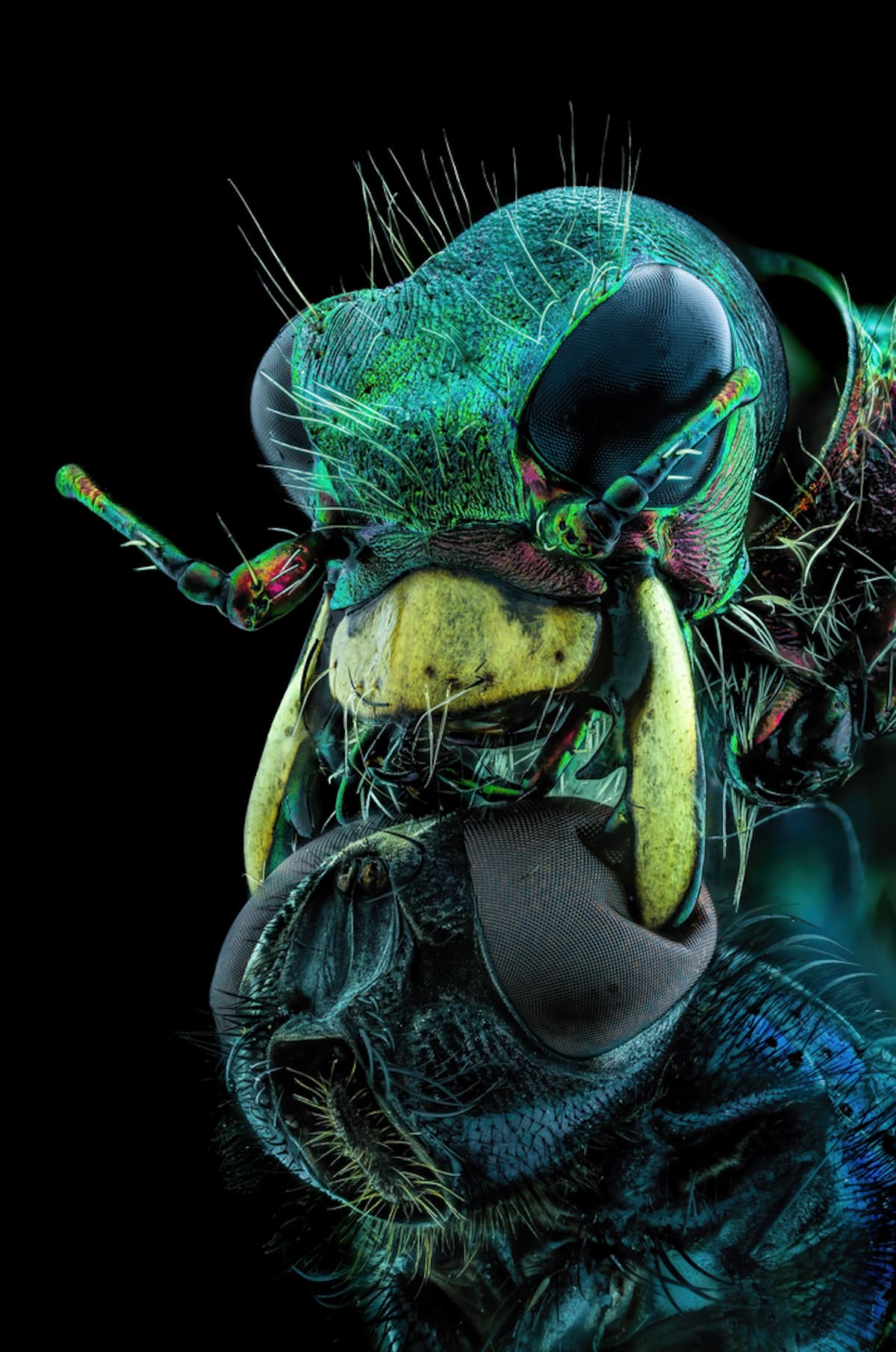
A fly under the chin of a tiger beetle, Murat Öztürk, 10th Place, Ankara, Turkey, Image Stacking, 3.7X (Objective Lens Magnification)
Unburned particles of carbon released when the hydrocarbon chain of candle wax breaks down, 6th Place, Ole Bielfeldt, Macrofying, Cologne, North Rhine-Westphalia, Germany, Brightfield, Image Stacking, 2.5X (Objective Lens Magnification)
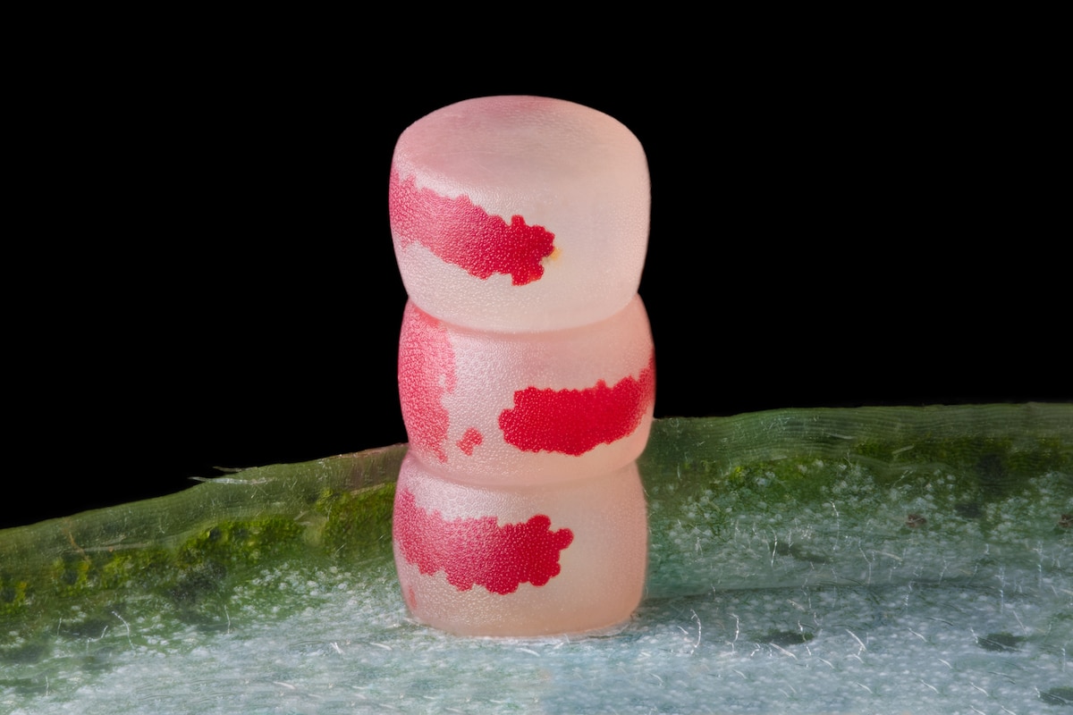
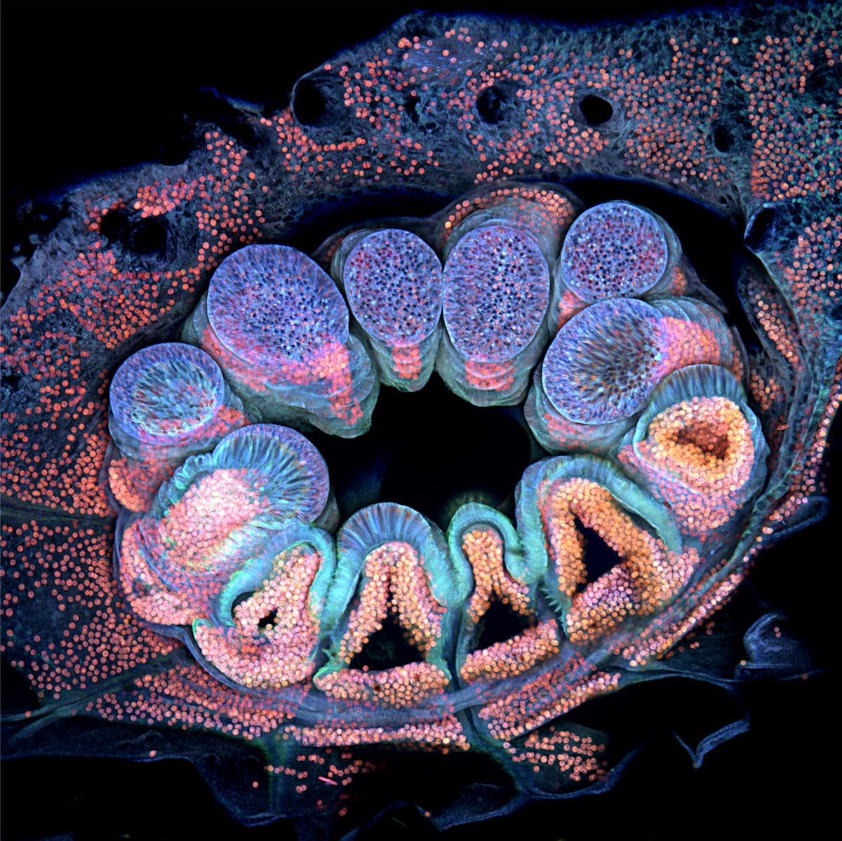
Moth eggs, Ye Fei Zhang, 11th Place, Jiang Yin, Jiangsu, China, Image Stacking, 10X (Objective Lens Magnification)
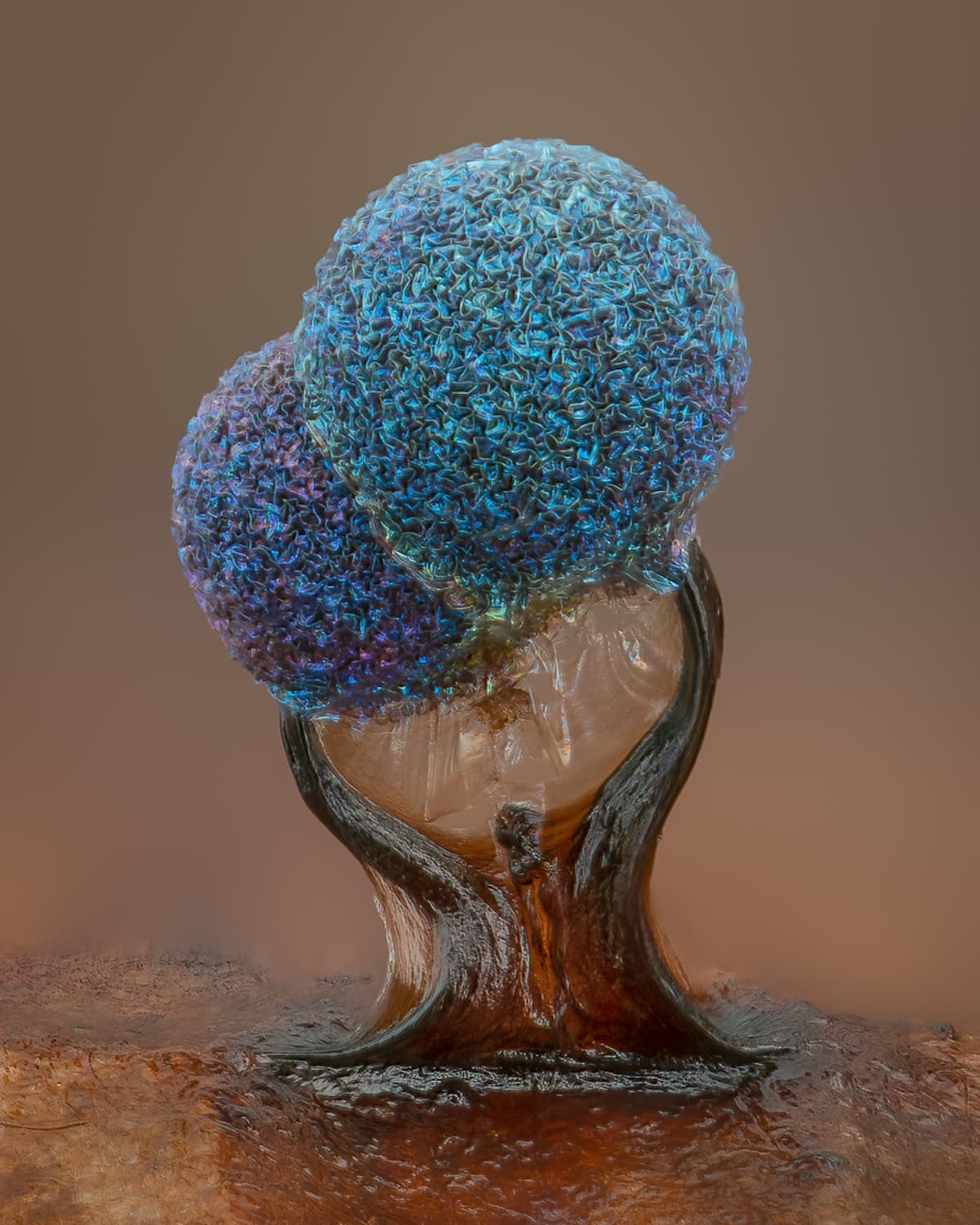
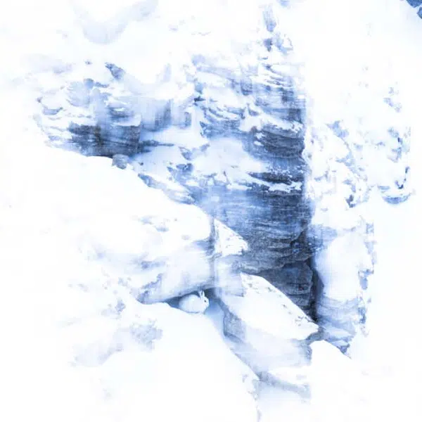
Slime mold (Lamproderma), 5th Place, Alison Pollack, San Anselmo, California, USA, Image Stacking, Reflected Light, 10X (Objective Lens Magnification)
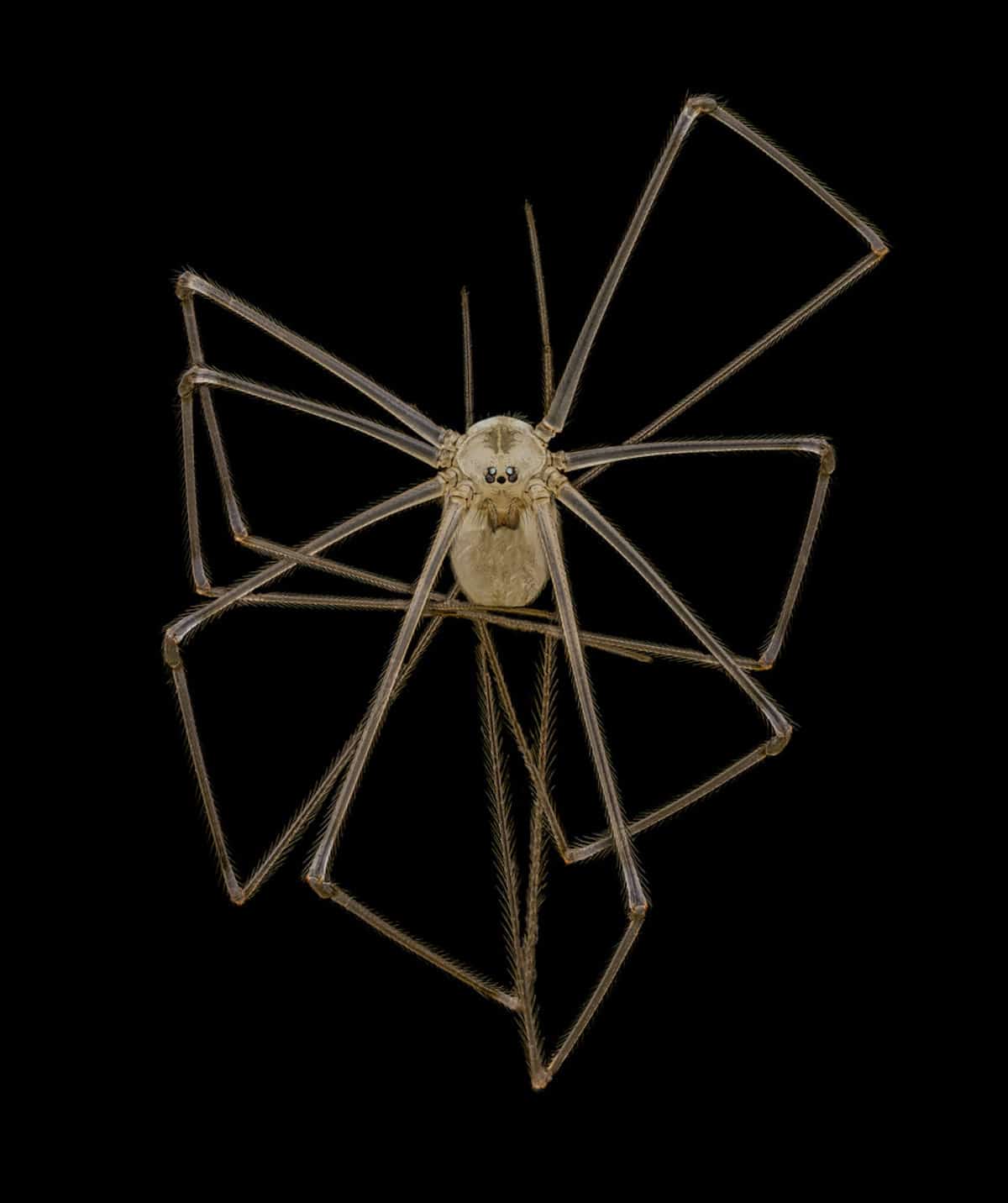
Long-bodied cellar/daddy long-legs spider (Pholcus phalangioides), 4th Place, Dr. Andrew Posselt, University of California, San Francisco (UCSF), Department of Surgery, Image Stacking, 3X (Objective Lens Magnification)
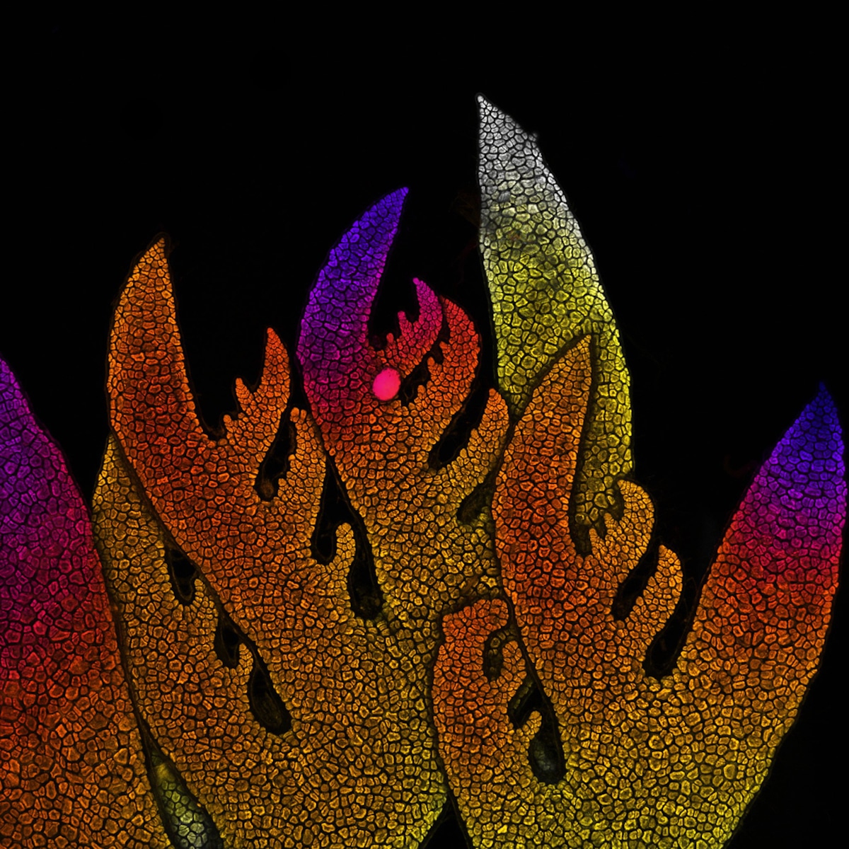
Growing tip of a red algae, Dr. Nathanaël Prunet, 8th Place, University of North Carolina at Chapel Hill, Department of Biology, Confocal, 10X (Objective Lens Magnification)
The 2022 contest received almost 1,300 entries from 72 countries.
Blood vessel networks in the intestine of an adult mouse, 3rd Place, Satu Paavonsalo & Dr. Sinem Karaman, University of Helsinki, Individualized Drug Therapy Research Program, Faculty of Medicine, Confocal, 10X (Object Lens Magnification)

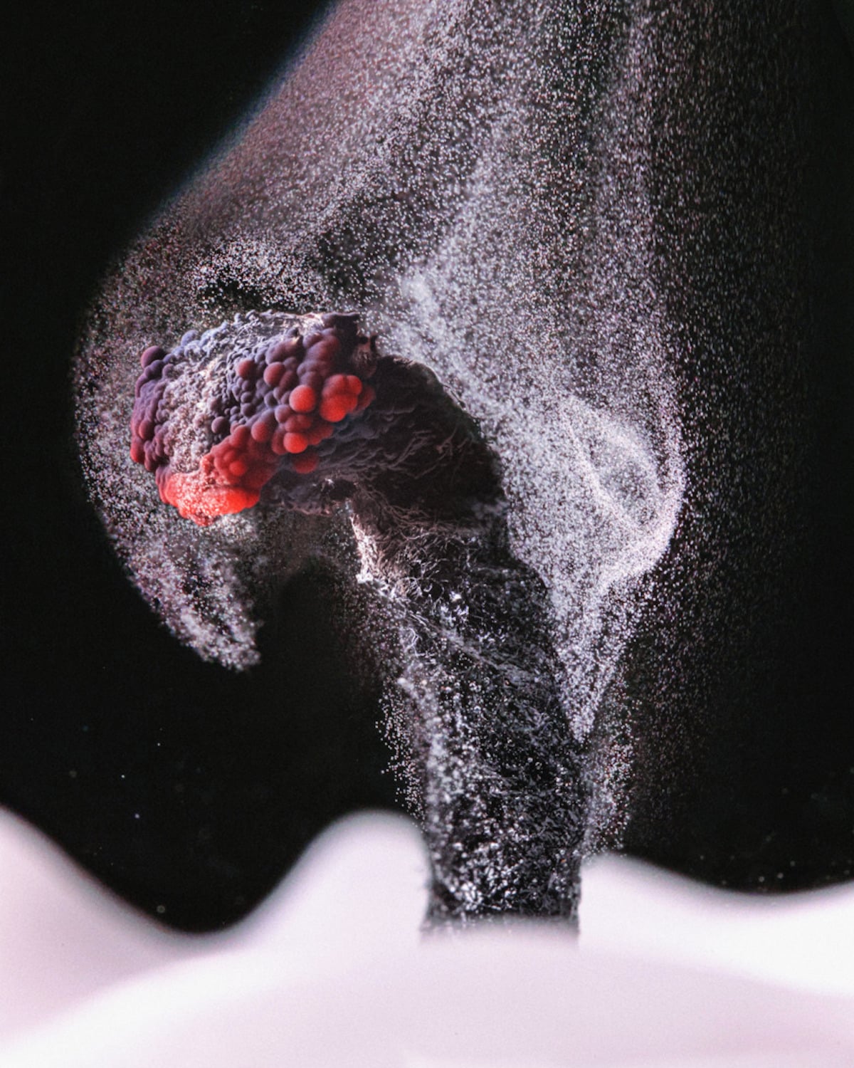
Breast tissue showing contractile myoepithelial cells wrapped around milk-producing alveoli, 2nd Place, Dr. Caleb Dawson, WEHI, The Walter and Eliza Hall Institute of Medical Research, Department of Immunology, Confocal, 40X (Objective Lens Magnification)

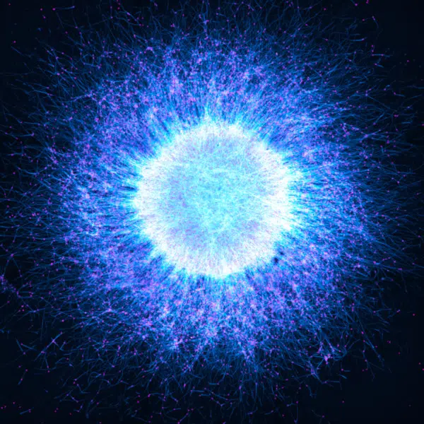
Human neurons derived from neural stem cells (NSCs), Dr. Jianqun Gao & Prof. Glenda Halliday, 7th Place, University of Sydney, Central Clinical School / Professor Glenda Halliday's Lab, Confocal, Fluorescence, 20X (Objective Lens Magnification)
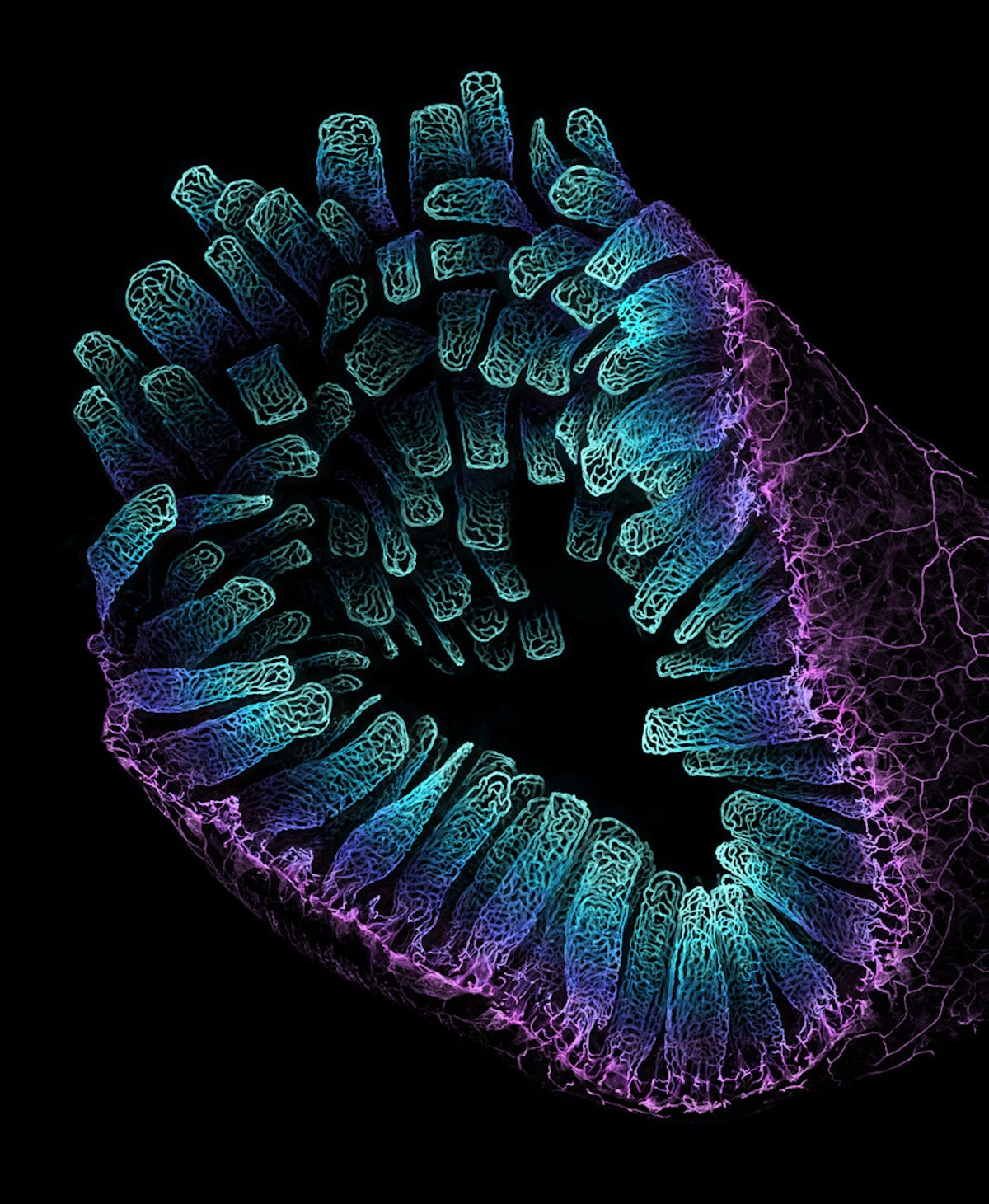
Autofluorescence of a single coral polyp (approx. 1 mm), Brett M. Lewis, 12th Place, Queensland University of Technology, Department of Earth and Atmospheric Science, Fluorescence, Image Stacking, 20X (Objective Lens Magnification)
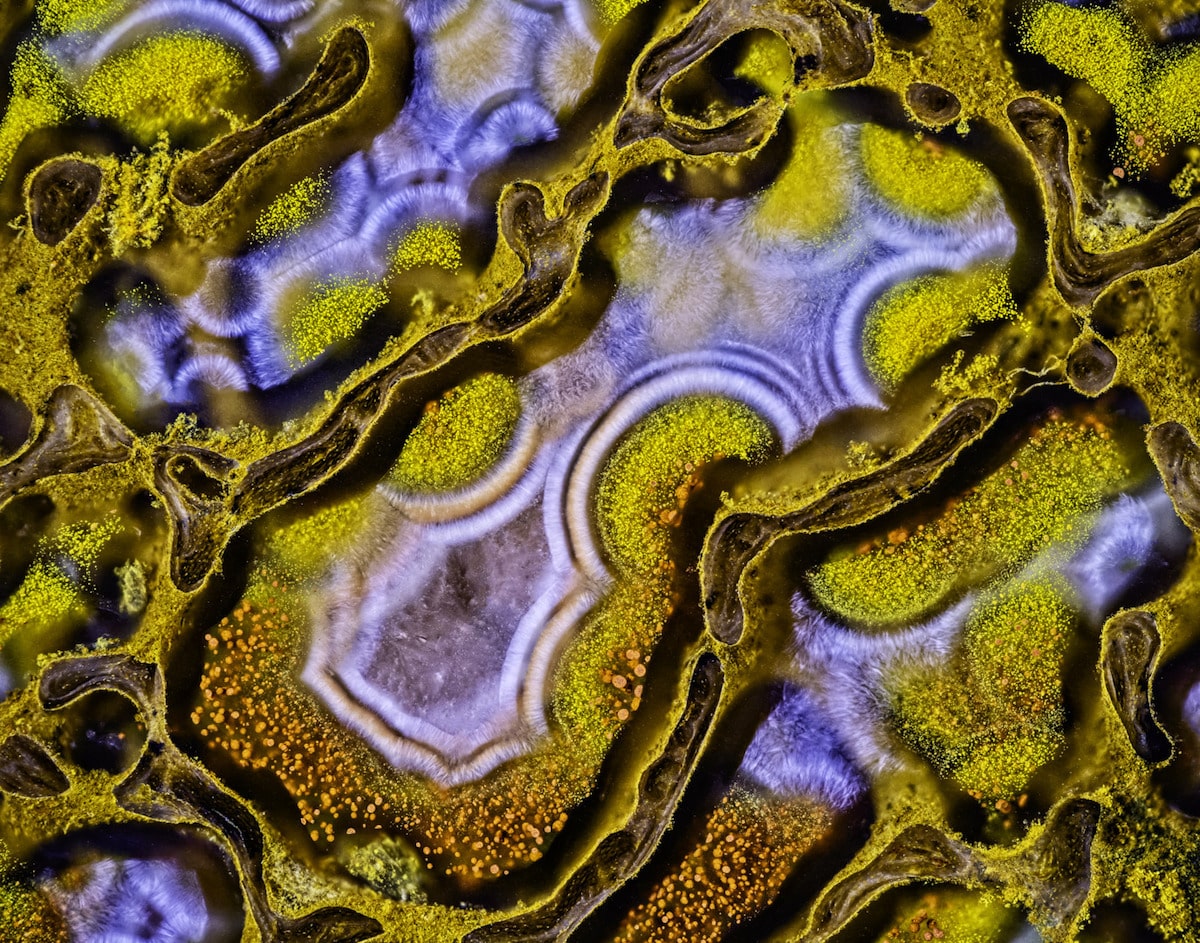
Agatized dinosaur bone, Randy Fullbright, 13th Place, Fullbright Studio, Vernal, Utah, USA, Image Stacking, 60X (Objective Lens Magnification)
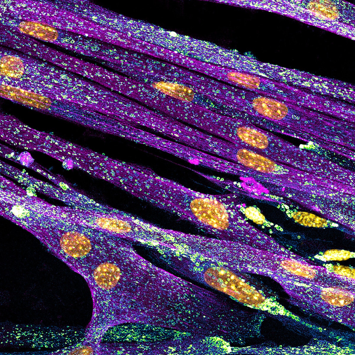
Differentiated cultured mouse myoblasts with lysosomes (cyan/green), nuclei (yellow), F-actin (magenta), Nadia Efimova, 14th Place, Amicus Therapeutics, Philadelphia, Pennsylvania, USA, Confocal, 40X (Objective Lens Magnification)
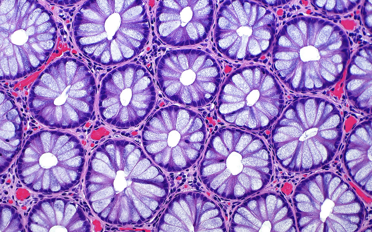
Cross sections of normal human colon epithelial crypts, Dr. Ziad El-Zaatari, 15th Place, Houston Methodist Hospital, Department of Pathology and Genomic Medicine, Brightfield, 20X (Objective Lens Magnification)