Trichome (white appendages) and stomata (purple pores) on a southern live oak leaf, 1st Place, Jason Kirk, Baylor College of Medicine, Optical Imaging & Vital Microscopy Core, Houston, Texas, USA, Image Stacking, 60X (Objective Lens Magnification)
The 47th annual Nikon Small World Photomicrography Competition has announced its winners and the results are as striking as ever. The contest celebrates the artistry of photography through a light microscope and, as usual, the top 20 winners photographed a wide variety of subjects using different methods. From confocal to fluorescence to polarized light, different types of microscopy were used to achieve these highly artistic images.
First place was awarded to Jason Kirk of the Baylor College of Medicine for his colorful image of a live oak leaf. By focusing on the appendages known as trichomes and the pores that allow the leaf to breathe, he's given an artistic and abstract take on a well-known piece of nature. To put the photo together, Kirk used a custom-made microscope system and stacked about 200 individual images to achieve the final look.
While Kirk is the director of the Optical Imaging & Vital Microscopy Core facility at Baylor, he also spends his personal time perfecting his photomicrography. “I've learned a lot from the scientific community, having spent 20 plus years in this field doing microscopy at a fairly high level,” he said. “But I've also learned a lot from the people in the hobbyist environment. Small World is a great combination of the two groups, and you don't often get an opportunity to see that.”
The rest of the top 20 images are a wonderful cross-section showing everything from the common snowflake and table salt to the neuron of a rat embryo and a cross-section of a mouse intestine. The top winners all came away with cash prizes and the acknowledgment that they are at the top of the photomicrography field.
See more winning images from the 2021 Nikon Small World Photomicrography Competition.
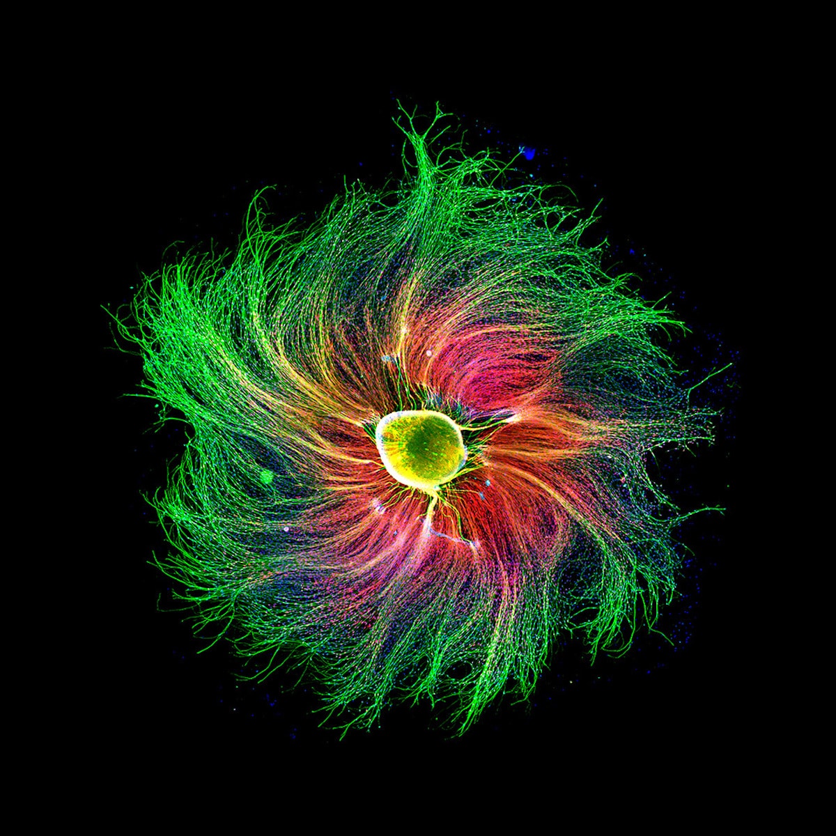
Sensory neuron from an embryonic rat, 4th Place, Paula Diaz, MinusPain, Pontificia Universidad Catolica de Chile, Department of Physiology, Santiago, Region Metropolitana, Chile, Fluorescence, 10X (Objective Lens Magnification)
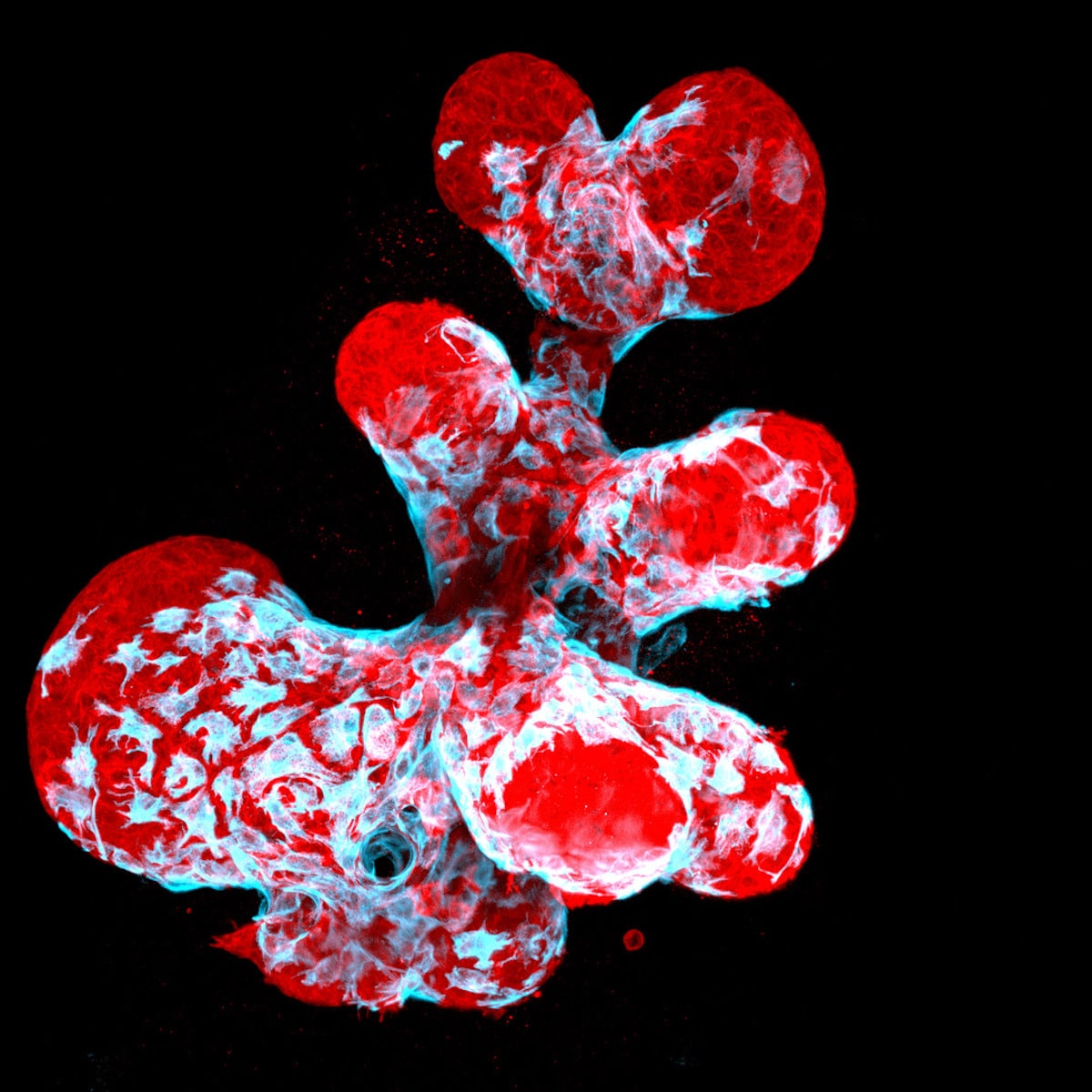
Breast organoid showing contractile myoepithelial cells (blue) crawling on secretory breast cells (red), 12th Place, Jakub Sumbal, Masaryk University, Department of Histology and Embryology, Brno, Czech Republic, Confocal, 40X (Objective Lens Magnification)
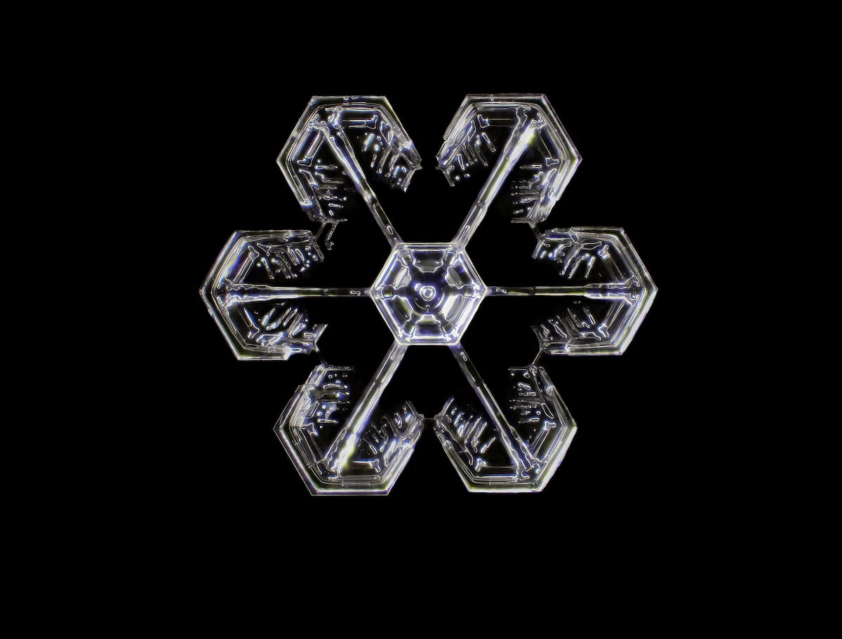
Snowflake, 14th Place, Dr. Joern N. Hopke, Waban, Massachusetts, USA, Image Stacking, 4X (Objective Lens Magnification)
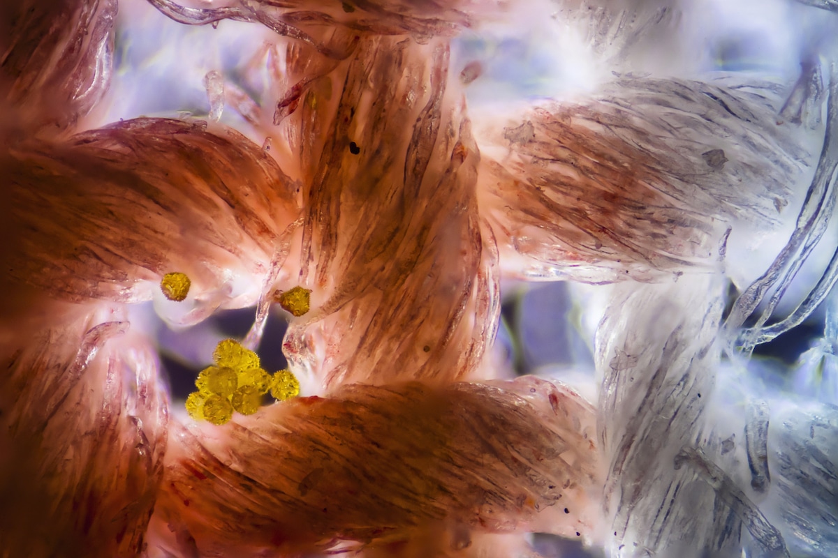
Cotton fabric with pollen grains, 13th Place, Dr. Felice Placenti, FP Nature and Landscape Photography, Siracusa, Sicilia, Italy, Darkfield, Image Stacking, 10X (Objective Lens Magnification)
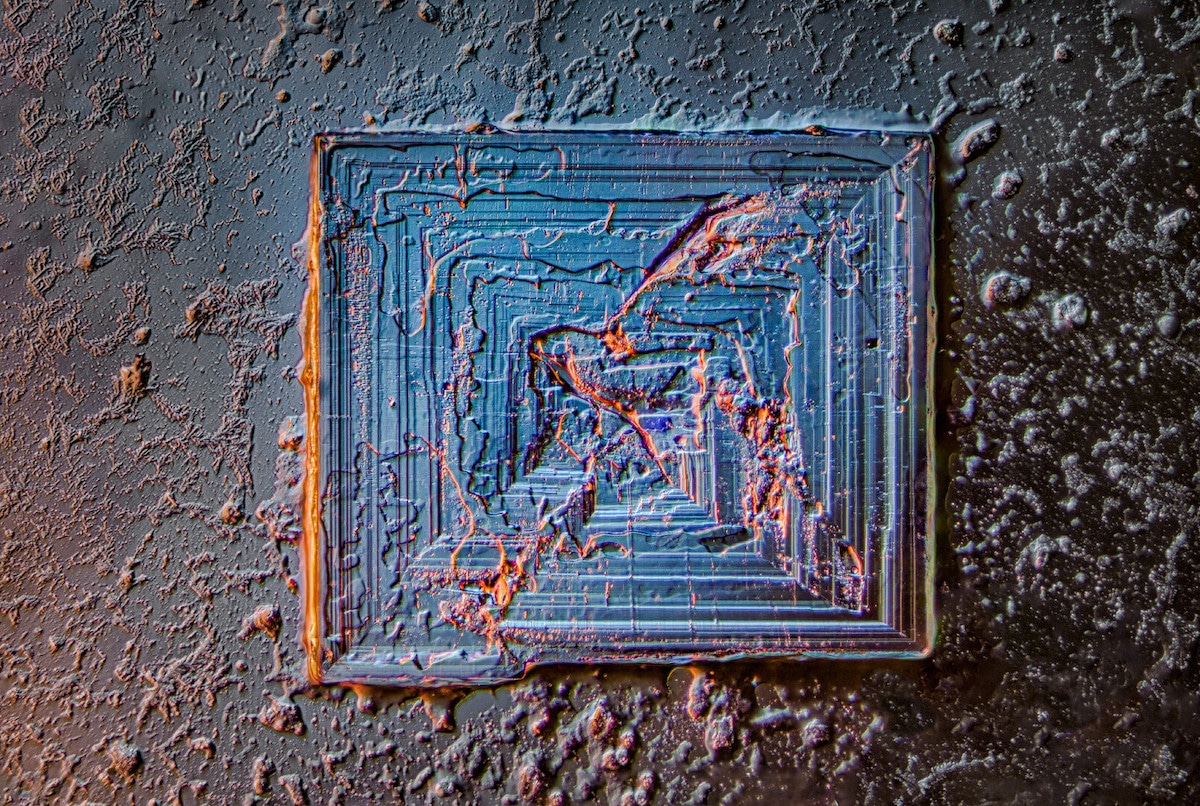
Table salt crystal, 18th Place, Saulius Gugis, Naperville, Illinois, USA, Image Stacking, Darkfield, Oblique, Rheinberg, Polarized Light, 10X (Objective Lens Magnification)
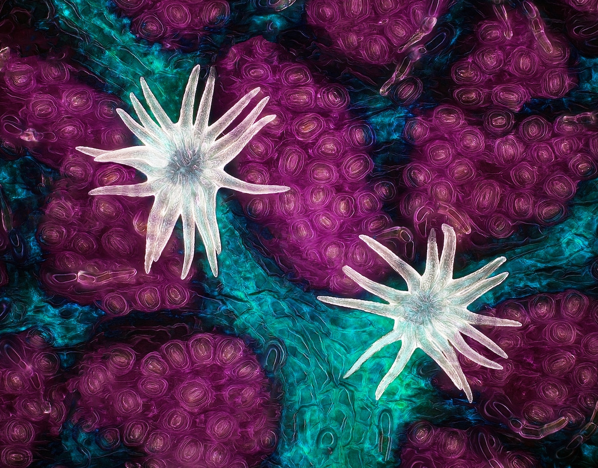
An in vivo snapshot of the neurons surrounding the mouth and tentacles of a juvenile starlet sea anemone (Nematostella vectensis), 16th Place, Ruohan Zhong, Stowers Institute for Medical Research, Gibson Lab,
Kansas City, Missouri, USA, Fluorescence, 20X (Objective Lens Magnification)
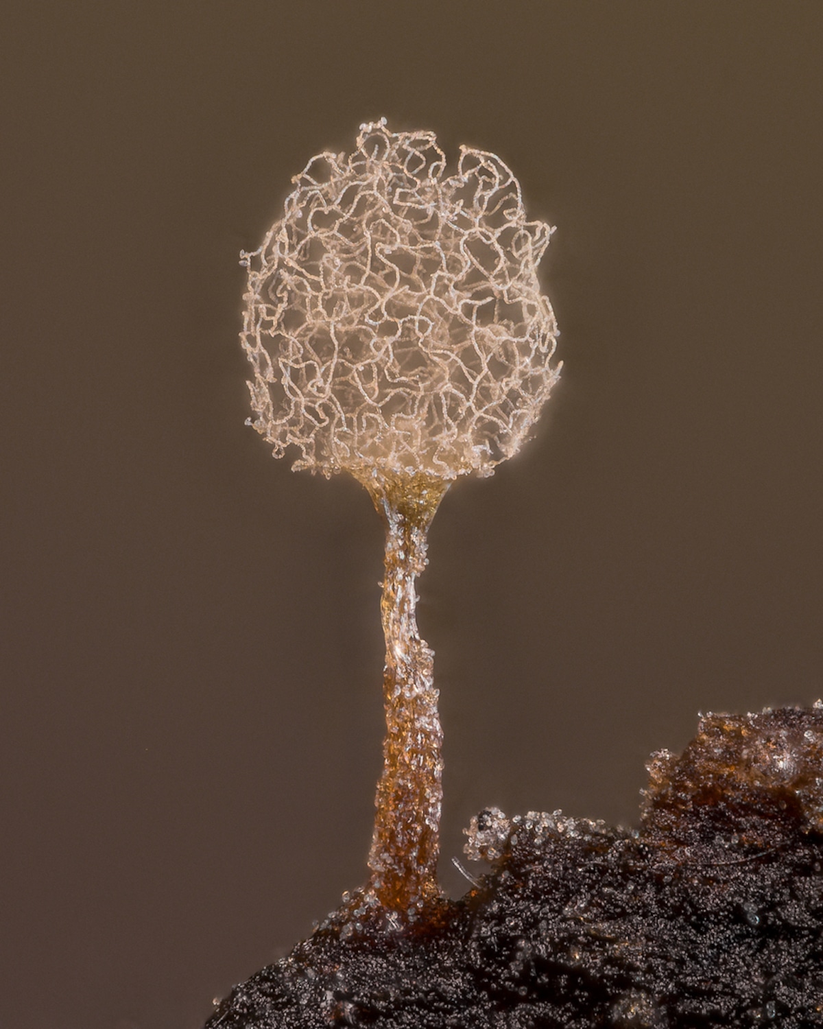
Slime mold (Arcyria pomiformis), 20th Place, Alison Pollack, San Anselmo, California, USA, Image Stacking, Reflected Light, 10X (Objective Lens Magnification)

Filamentous strands of Nostoc cyanobacteria captured inside a gelatinous matrix, 17th Place, Martin Kaae Kristiansen, My Microscopic World, Aalborg, Nordjylland, Denmark, Image Stacking, Polarized Light, 4X (Objective Lens Magnification)

Proboscis of a housefly (Musca domestica), 5th Place, Oliver Dum, Medienbunker Produktion, Bendorf, Rheinland Pfalz, Germany, Image Stacking, 40X (Objective Lens Magnification)
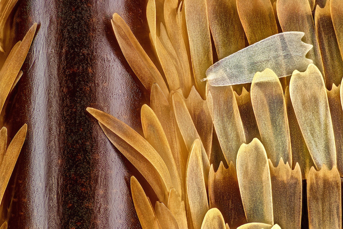
Vein and scales on a butterfly wing (Morpho didius), 10th Place, Sébastien Malo, Saint Lys, Haute-Garonne, France, Image Stacking, Reflected Light, 20X (Objective Lens Magnification)

Calcite crystal inclusion suspended in a spinel gemstone, 19th Place, Billie Hughes, Lotus Gemology, Bangkok, Thailand, Darkfield, Image Stacking, 40X (Objective Lens Magnification)
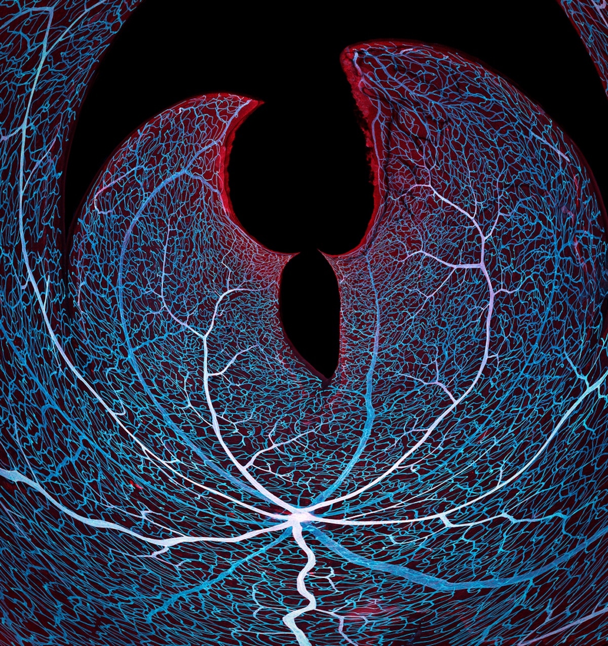
Vasculature of a mouse retina, 11th Place, Jason Kirk & Carlos P. Flores Suarez, Baylor College of Medicine, Optical Imaging & Vital Microscopy Core, Houston, Texas, USA, Confocal, 20X (Objective Lens Magnification)
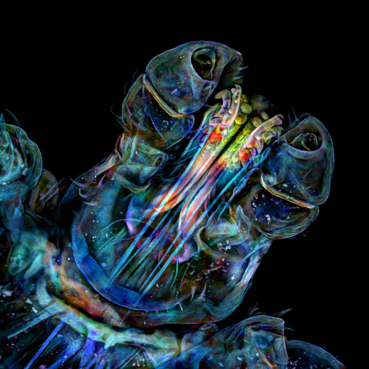
Head of a tick, 7th Place, Dr. Tong Zhang & Dr. Paul Stoodley, The Ohio State University, Campus Microscopy & Imaging Facility, Columbus, Ohio, USA, Confocal, 10X (Objective Lens Magnification)
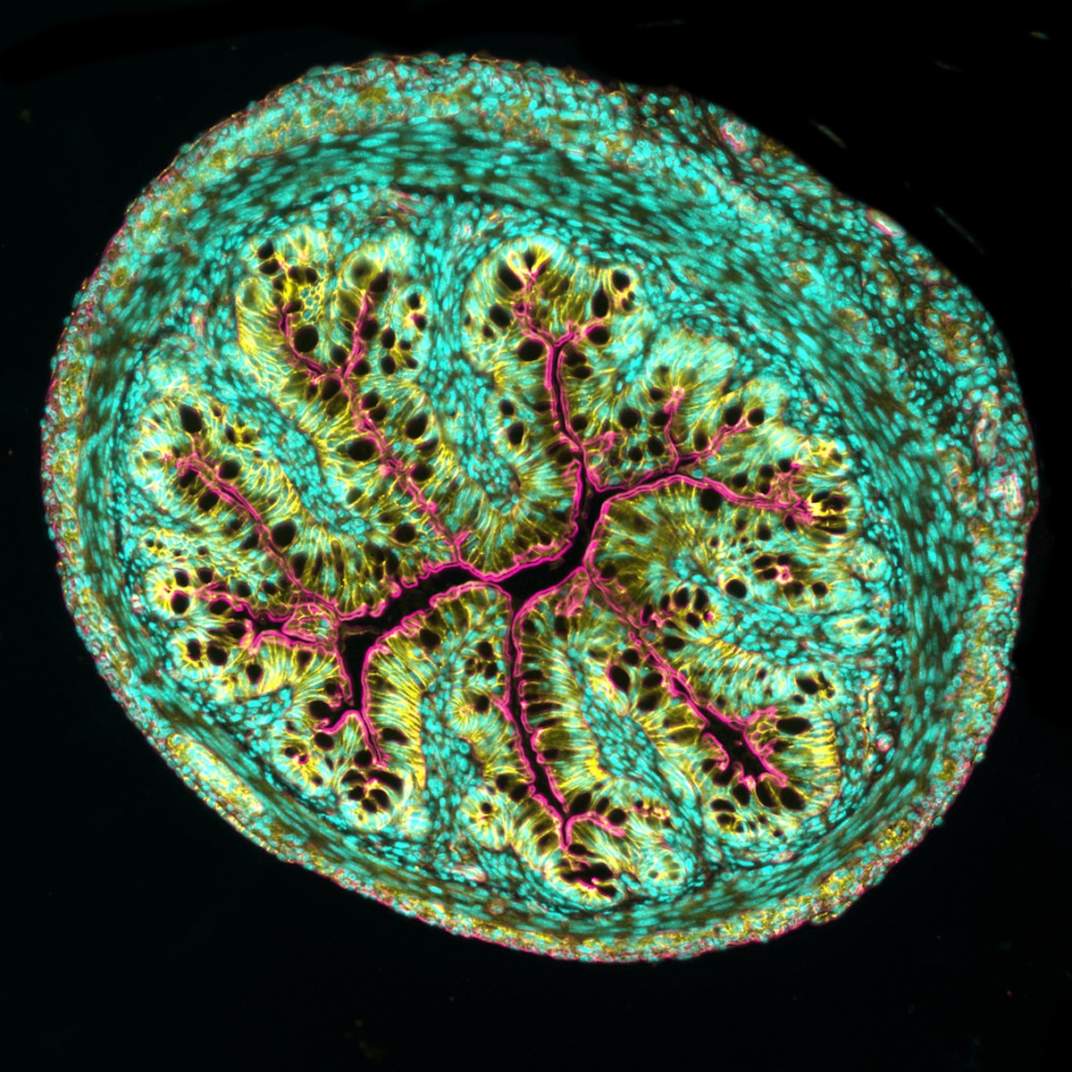
Cross section of mouse intestine, 8th Place, Dr. Amy Engevik, Medical University of South Carolina, Department of Regenerative Medicine and Cell Biology, Charleston, South Carolina, USA, Fluorescence, 10X (Objective Lens Magnification)
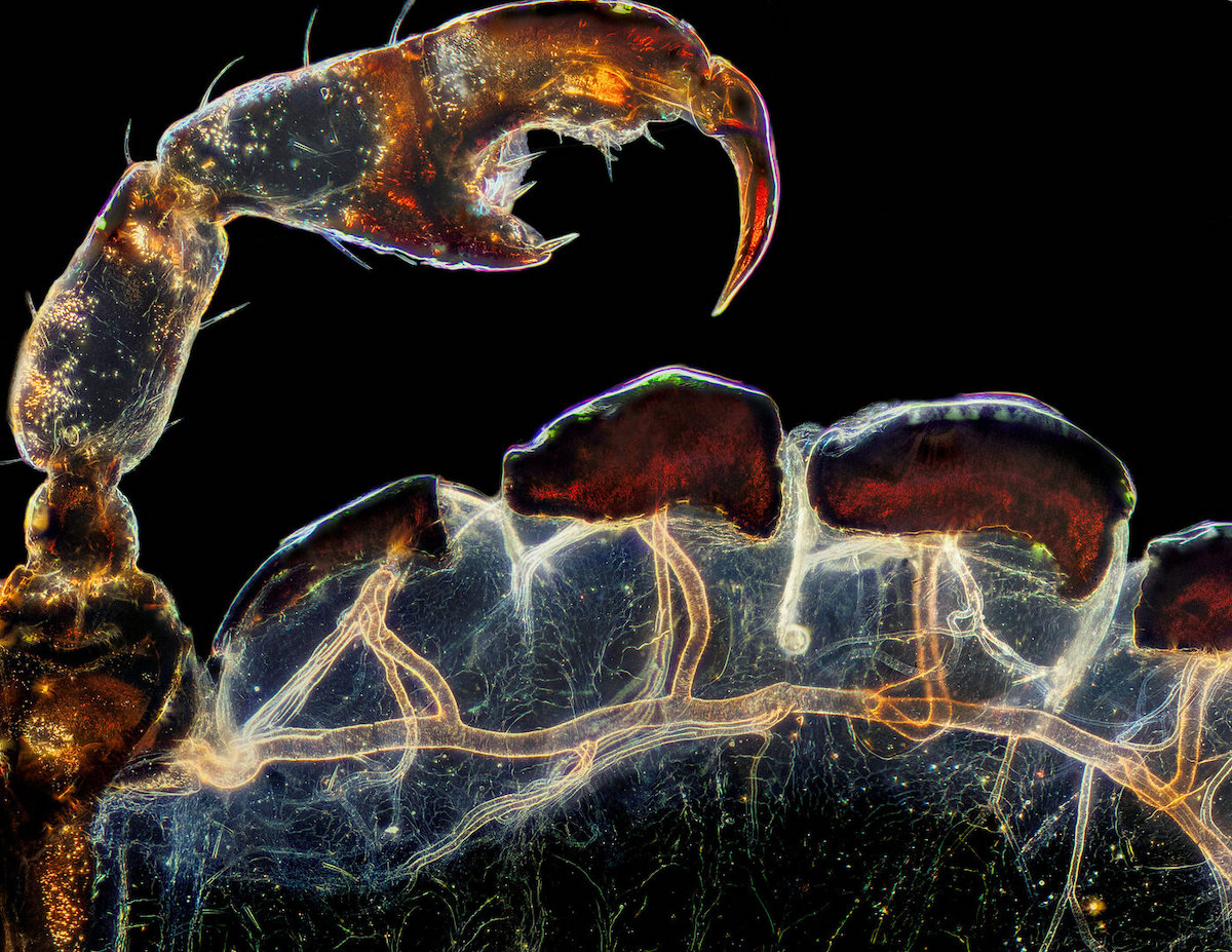
Rear leg, claw, and respiratory trachea of a louse (Haematopinus suis), 3rd Place, Frank Reiser, Nassau Community College, Department of Biology, Garden City, New York, USA, Darkfield, Image Stacking, 5X (Objective Lens Magnification)
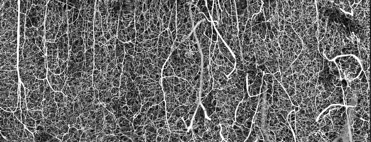
3D vasculature of an adult mouse brain (somatosensory cortex) , 6th Place, Dr. Andrea Tedeschi, The Ohio State University / Wexner Medical Center Department of Neuroscience, Columbus, Ohio, USA, Confocal, 10X (Objective Lens Magnification)

Water flea (Daphnia), carrying embryos and peritrichs, 9th Place, Jan van IJken, Jan van IJken Photography and Film
Amsterdam, Noord-Holland, Netherlands, Darkfield, Image Stacking, 10X (Objective Lens Magnification)
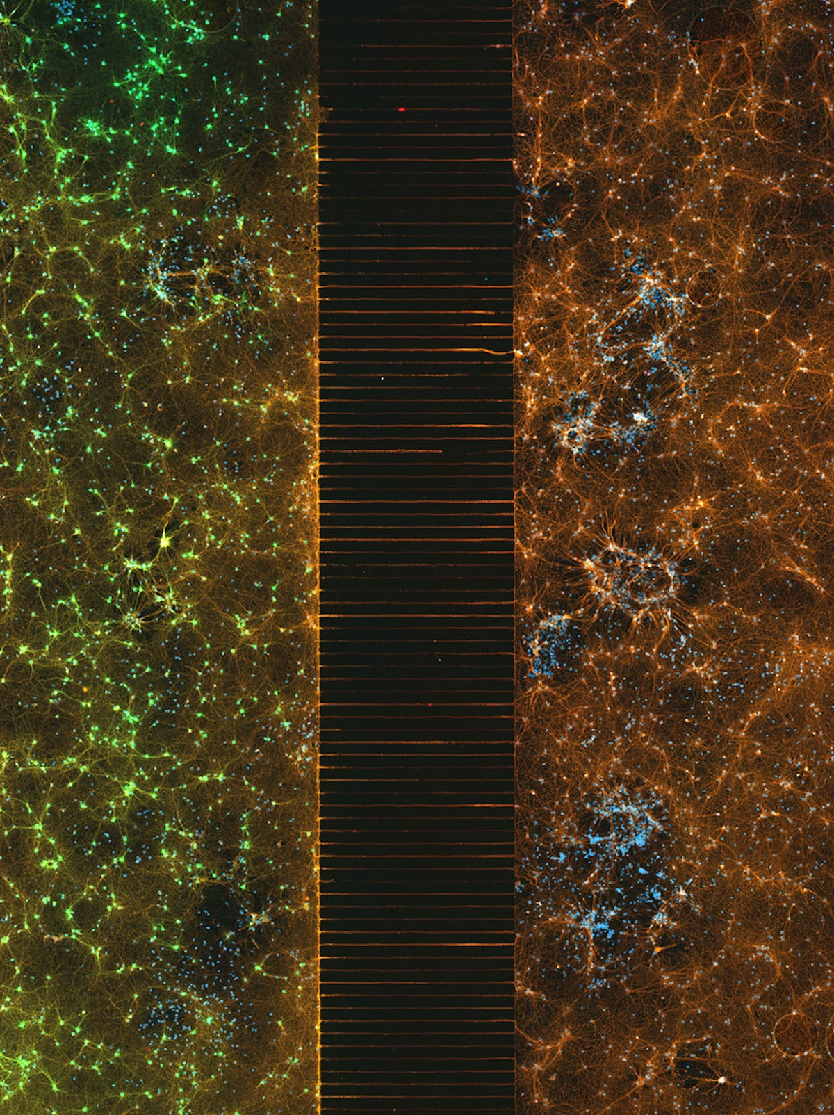
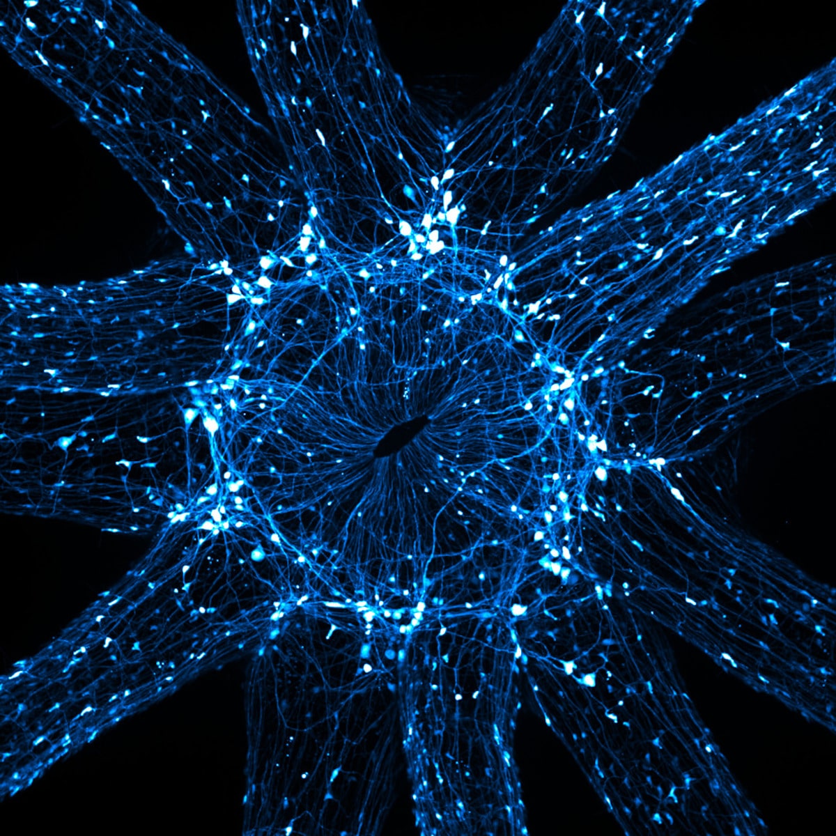
A microfluidic device containing 300k networking neurons in 2 isolated populations. Both sides were treated with a unique virus and bridged by axons, 2nd Place, Esmeralda Paric & Holly Stefen, Dementia Research Centre, Macquarie University
Department of Biomedical Sciences,
Macquarie Park, NSW, Australia, Fluorescence, 40X (Objective Lens Magnification)
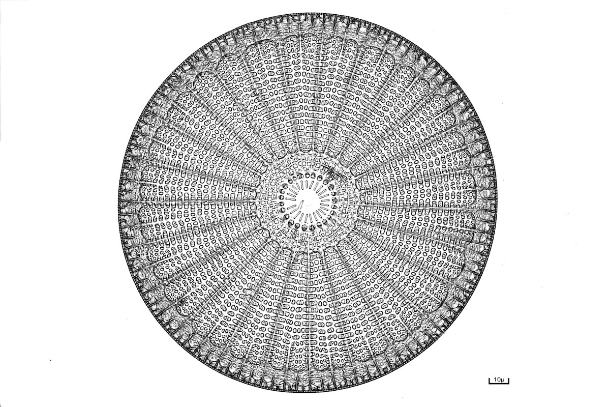
Diatom (Arachnoidiscus), 15th Place, Bernard Allard, Club Français de Microscopie, Sucy-en-Bry, France, Brightfield, Image Stacking, 40X (Objective Lens Magnification)