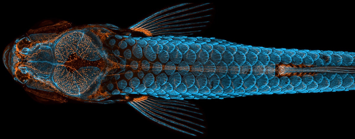
Dorsal view of bones and scales (blue) and lymphatic vessels (orange) in a juvenile zebrafish. Daniel Castranova, Dr. Brant Weinstein & Bakary Samasa (Eunice Kennedy Shriver National Institute of Child Health and Human Development, National Institutes of Health, Bethesda, Maryland, USA). First Place. Confocal, 4X (Objective Lens Magnification).
Nikon's celebration of life under the microscope is back. Since 1974, the Nikon Small World Photomicrography Competition has highlighted the artistry that scientists, researchers, and enthusiasts can create. By using their skills and creativity, the participants open up a whole new world to the public. This year's contest was as thrilling as ever, with a group of scientists from the National Institute of Health taking home the top prize.
Daniel Castranova, assisted by Bakary Samasa while working in the lab of Dr. Brant Weinstein, captured the winning image of a young zebrafish. With the bones and scales tinged blue and the lymphatic vessels colored orange, the photograph gives an informative and aesthetically pleasing look at this fish's anatomy. The image is not only beautiful but groundbreaking, as it helped the team see that zebrafish actually have lymphatic vessels in their skull. This was previously only thought to occur in mammals.
“The image is beautiful, but also shows how powerful the zebrafish can be as a model for the development of lymphatic vessels,” Castranova said, “Until now, we thought this type of lymphatic system associated with the nervous system only occurred in mammals. By studying them, the scientific community can expedite a range of research and clinical innovations–everything from drug trials to cancer treatments. This is because fish are so much easier to raise and image than mammals.”
Over 2,000 examples of photomicrography were entered into this year's competition. Submissions were gathered from scientists, hobbyists, and artists from 90 countries. An expert panel of judges, which included a photo editor from National Geographic, a well as a representative from NASA, culled this list into their top 20 favorites. Take a look at the top photographs and, if you like what you see, keep checking back to the Nikon Small World website to see when their full-color calendar of this year's winners will be released.
Look at life under the microscope thanks to the winners of the 2020 Nikon Small World Photomicrography Contest.
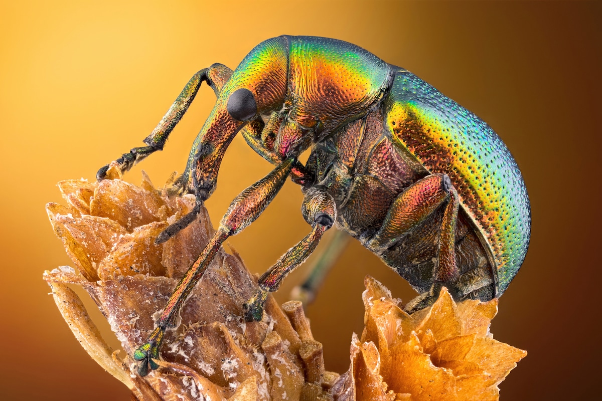
Leaf roller weevil (Byctiscus betulae) lateral view. Özgür Kerem Bulur (Istanbul, Turkey). Fourteenth Place. Image Stacking, Reflected Light, 3.7X (Objective Lens Magnification).
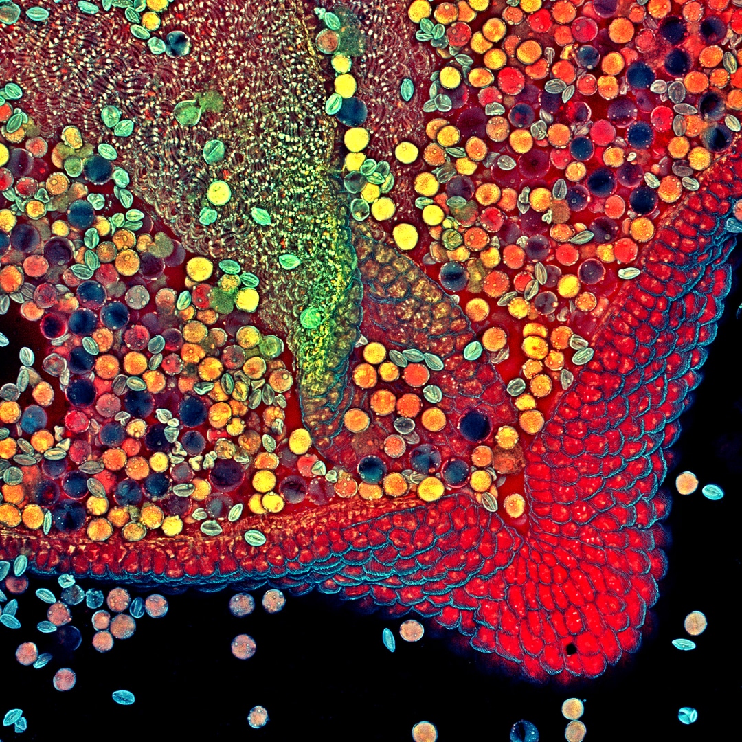
Hebe plant anther with pollen. Dr. Robert Markus & Zsuzsa Markus (University of Nottingham, School of Life Sciences, Super Resolution Microscopy, Nottingham, United Kingdom). Sixth Place. Confocal, 10X (Objective Lens Magnification).
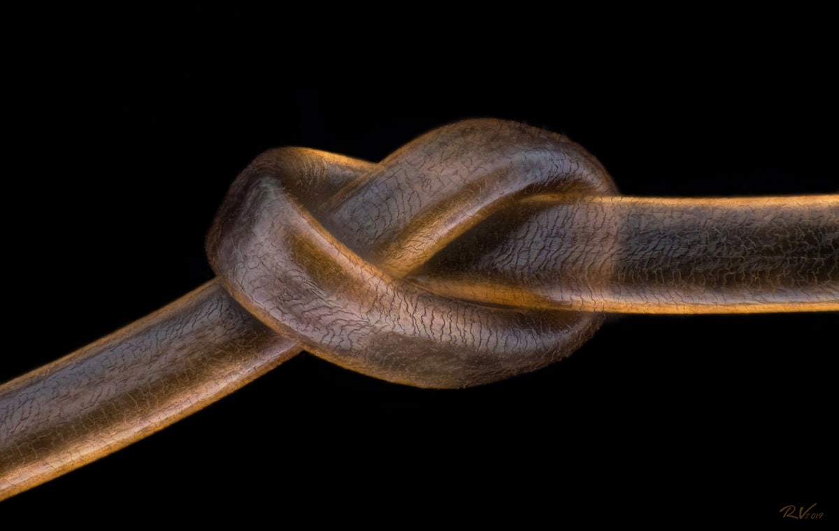
Human hair. Robert Vierthaler (Pfarrwerfen, Salzburg, Austria). Twelfth Place. Image Stacking, 20X (Objective Lens Magnification).

Bogong moth. Ahmad Fauzan (Saipem, Jakarta, Indonesia). Fifth Place. Image Stacking, 5X (Objective Lens Magnification).
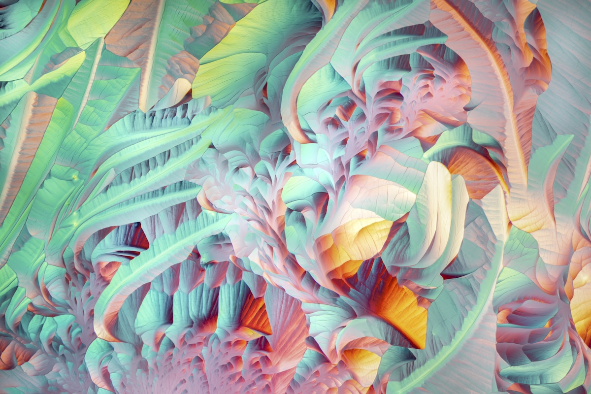
Crystals formed after heating an ethanol and water solution containing L-glutamine and beta-alanine. Justin Zoll (Ithaca, New York, USA). Thirteenth Place. Polarized Light, 4X (Objective Lens Magnification).
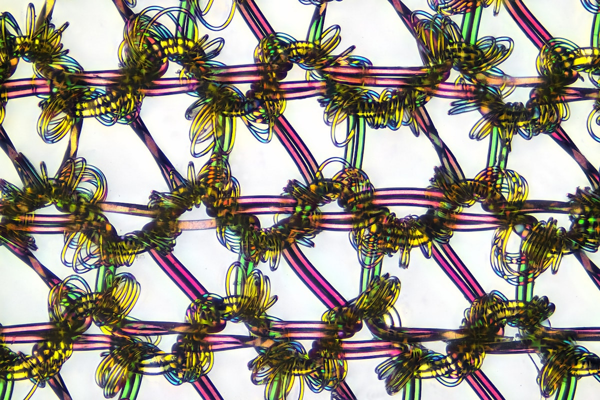
Nylon stockings. Alexander Klepnev (JSC Radiophysics, Moscow, Russian Federation). Sixteenth Place. Polarized Light, 9X (Objective Lens Magnification).
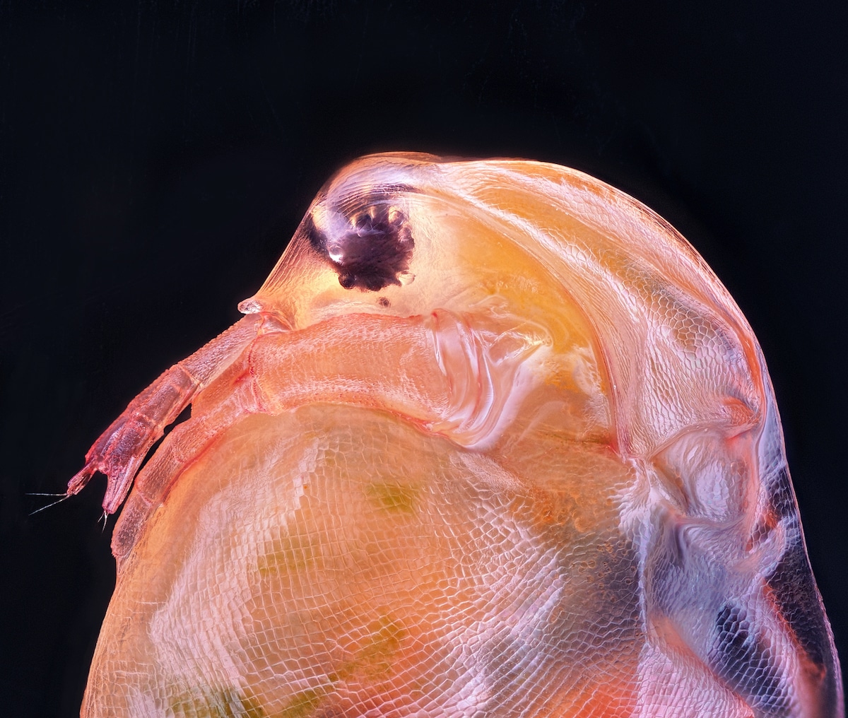
Daphnia magna (Phyllopoda). Ahmad Fauzan (Saipem, Jakarta, Indonesia). Tenth Place. Image Stacking, 10X (Objective Lens Magnification).
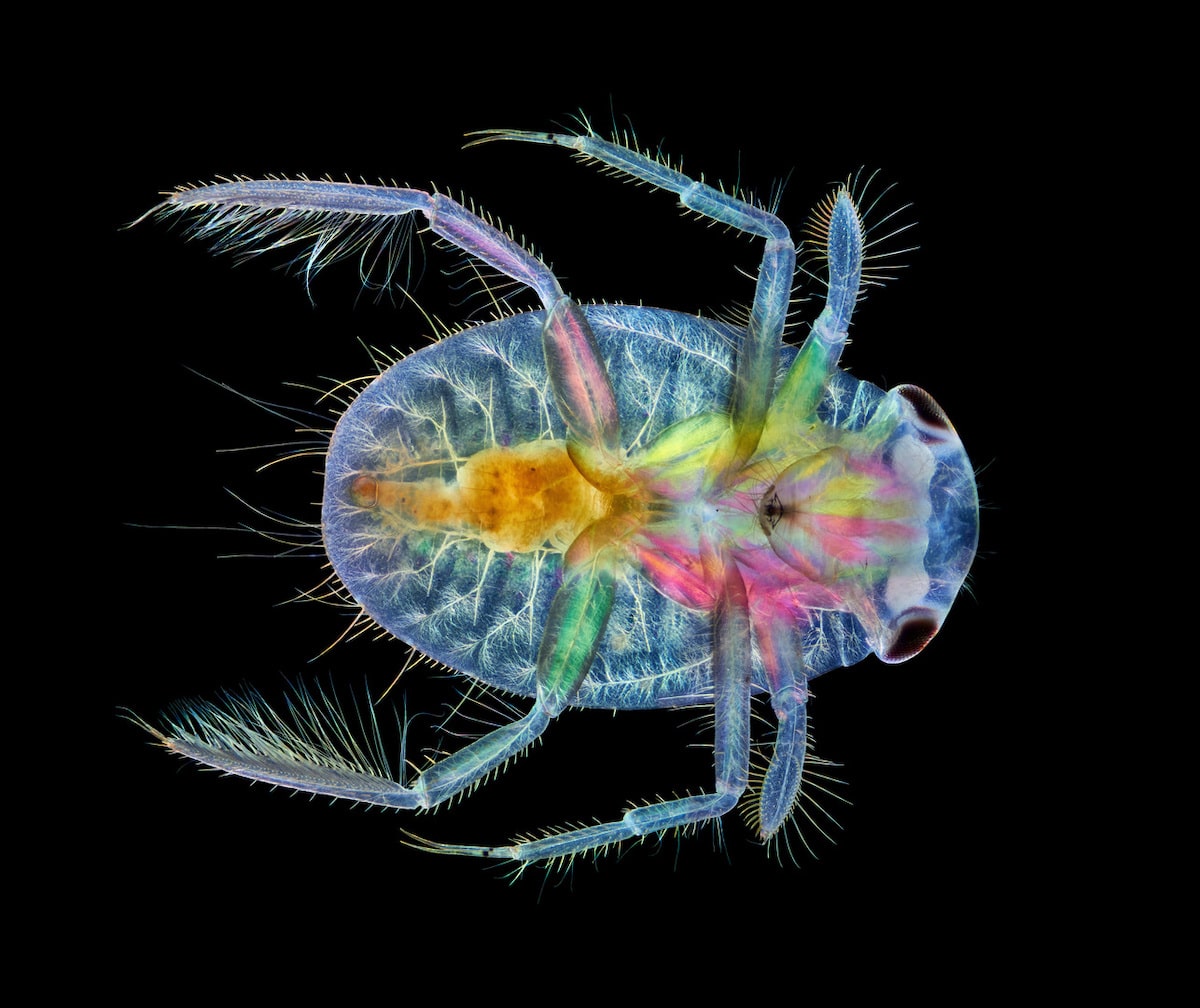
Ventral view of an immature water boatman. Anne Algar (Hounslow, Middlesex, United Kingdom). Seventeenth Place. Darkfield, Image Stacking, Polarized Light, 4X (Objective Lens Magnification).
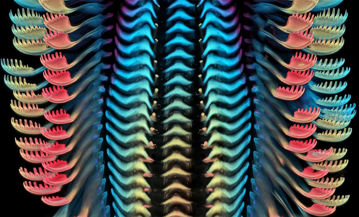
Tongue (radula) of a freshwater snail. Dr. Igor Siwanowicz (Howard Hughes Medical Institute, Ashburn, Virginia, USA). Third Place. Confocal, 40X (Objective Lens Magnification).
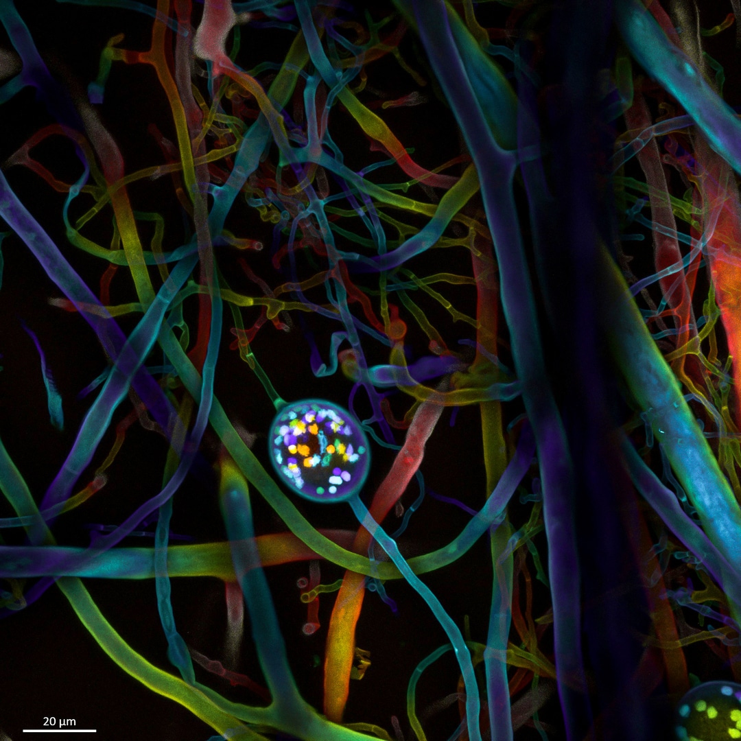
Multi-nucleate spores and hyphae of a soil fungus (arbuscular mycorrhizal fungus). Dr. Vasileios Kokkoris, Dr. Franck Stefani & Dr. Nicolas Corradi (University of Ottawa & Agriculture and Agrifood Canada, Ottawa, Ontario, Canada). Fourth Place. Confocal, 63X (Objective Lens Magnification).
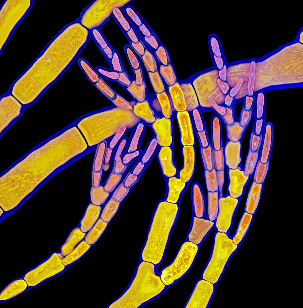
Red algae. Dr. Tagide deCarvalho (University of Maryland, Baltimore County, Baltimore, Maryland, USA). Eleventh Place. Confocal, 63X (Objective Lens Magnification).

Atlas moth wing. Chris Perani (San Rafael, California, USA). Eighteenth Place. Image Stacking, 10x (Objective Lens Magnification).
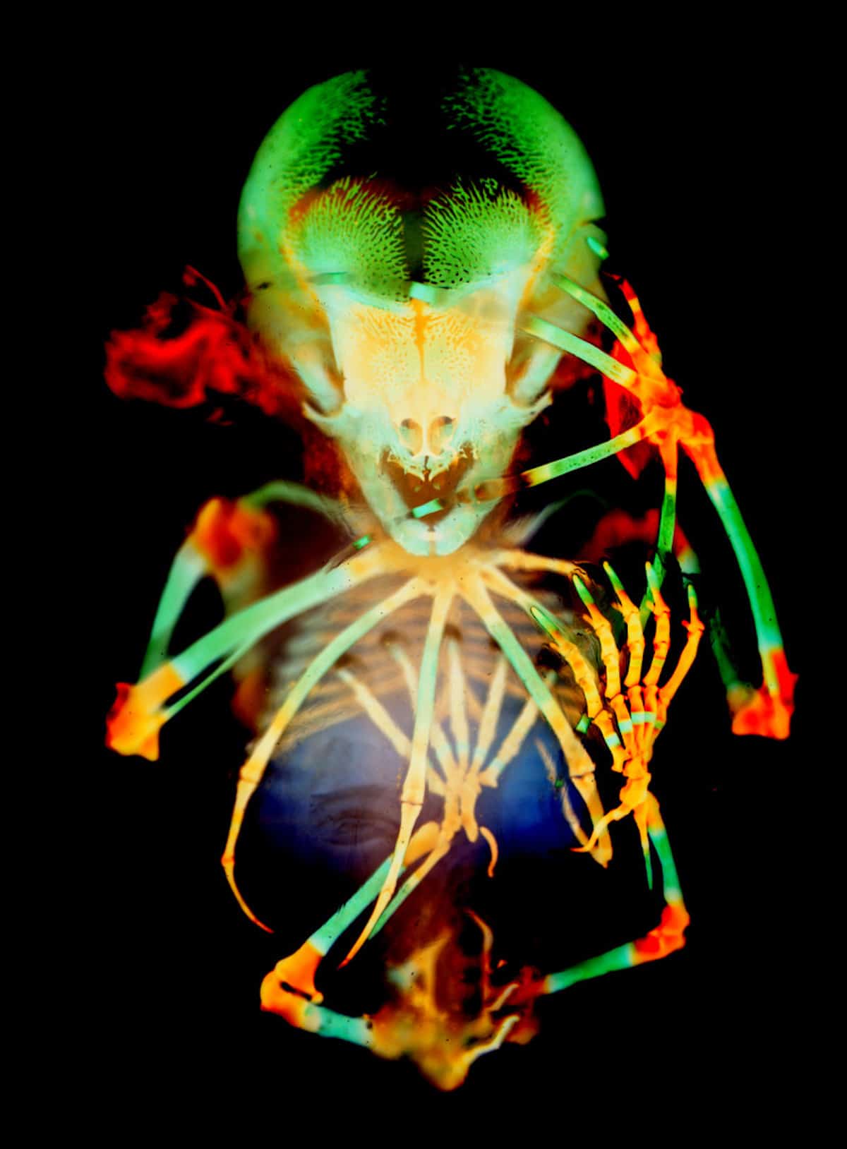
Skeleton preparation of a short-tailed fruit bat embryo (Carollia perspicillata). Dr. Dorit Hockman & Dr. Vanessa Chong-Morrison (University of Cape Town, Rondebosch, Cape Town, South Africa). Twentieth Place. Brightfield, 1X (Objective Lens Magnification).
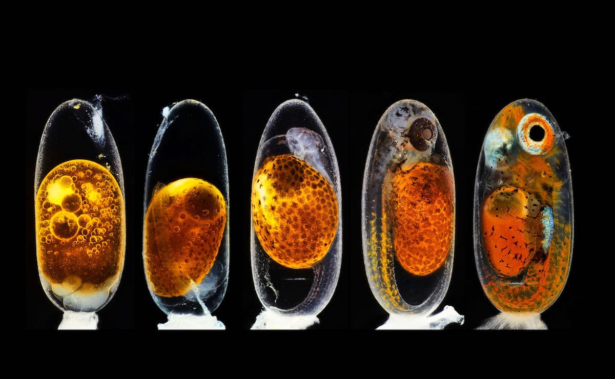
Embryonic development of a clownfish (Amphiprion percula) on days 1, 3 (morning and evening), 5, and 9. Daniel Knop (Natur und Tier-Verlag NTV, Oberzent-Airlenbach, Hessen, Germany). Second Place. Image Stacking, 10X (Objective Lens Magnification).

Microtubules (orange) inside a cell. Nucleus is shown in cyan. Jason Kirk (Baylor College of Medicine, Optical Imaging & Vital Microscopy Core, Houston, Texas, USA). Seventh Place. Confocal, 63X (Objective Lens Magnification).
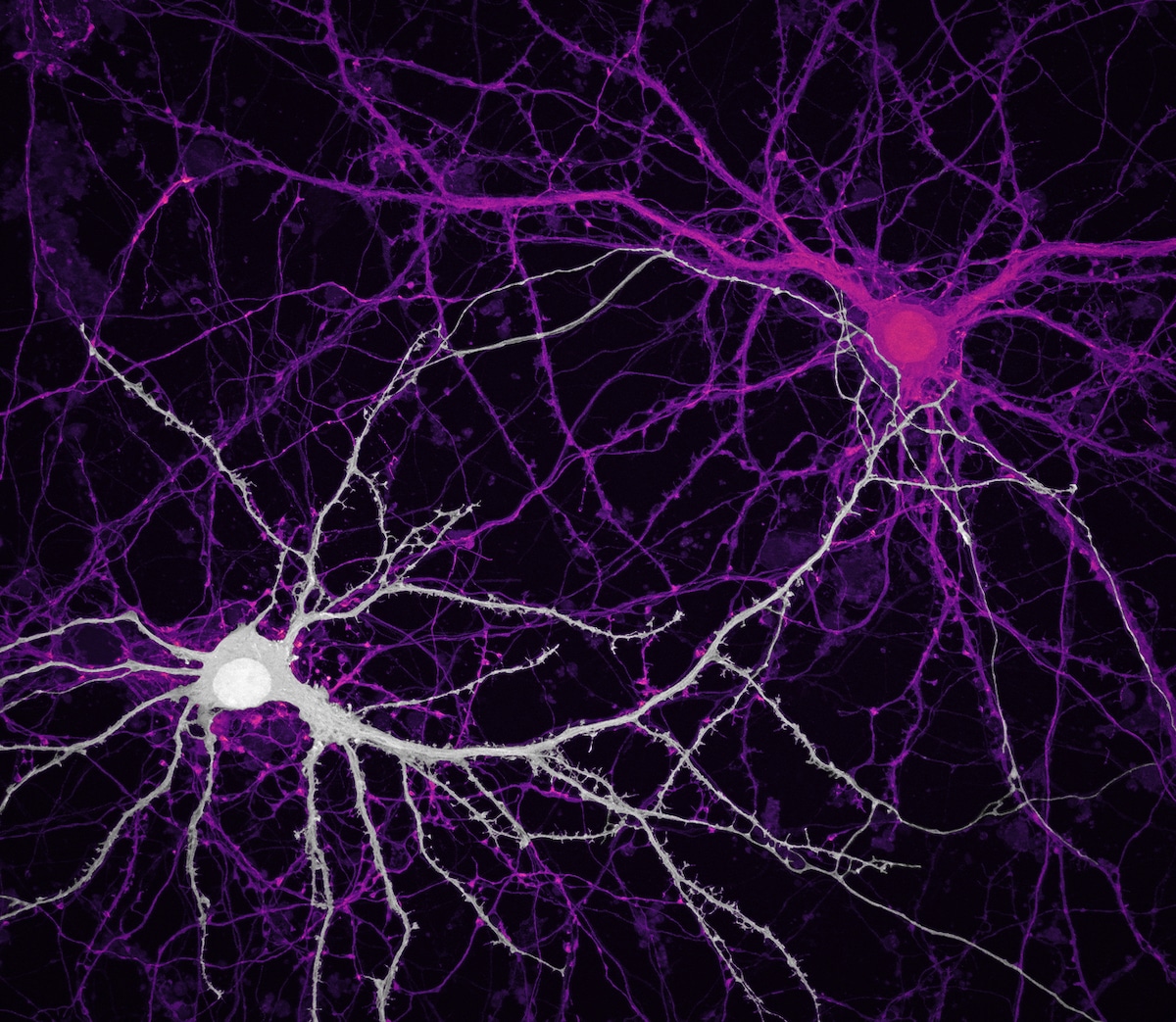
Connections between hippocampal neurons (brain cells). Jason Kirk & Quynh Nguyen (Baylor College of Medicine, Optical Imaging & Vital Microscopy Core, Houston, Texas, USA). Ninth Place. Confocal, 63X (Objective Lens Magnification).
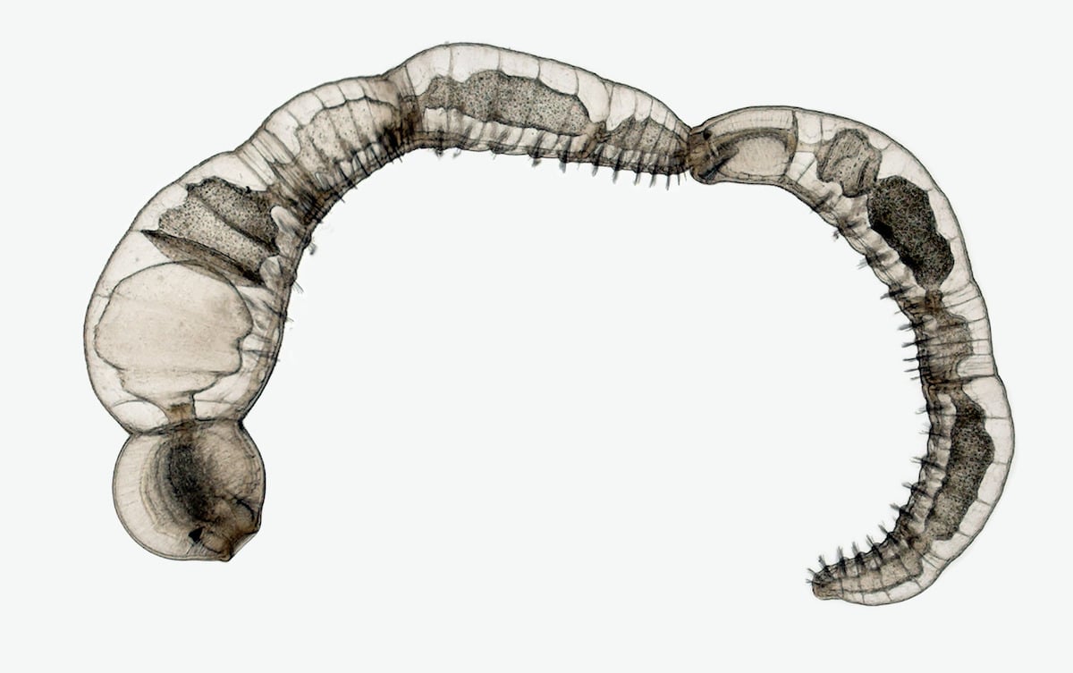
Chain of daughter individuals from the asexually reproducing annelid species Chaetogaster diaphanus. Dr. Eduardo Zattara & Dr. Alexa Bely (CONICET, Instituto Nac. de Investigaciones en Biodiversidad y Medio Ambiente, Bariloche, Rio Negro, Argentina). Fifteenth Place. Brightfield, 5X (Objective Lens Magnification).
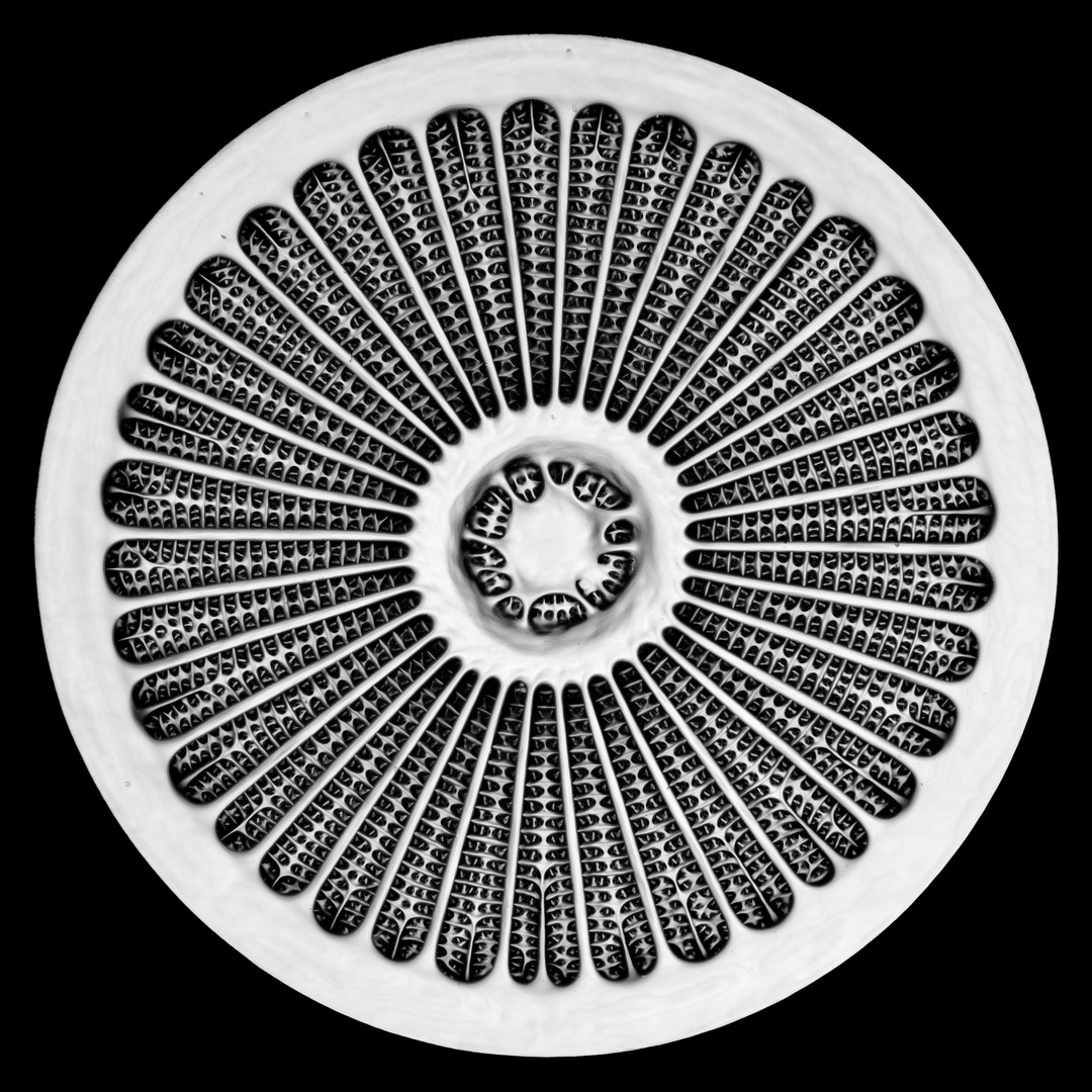
Silica cell wall of the marine diatom Arachnoidiscus sp. Dr. Jan Michels (Christian-Albrechts-Universität zu Kiel, Department of Functional Morphology and Biomechanics, Kiel, Schleswig-Holstein, Germany). Ninteenth Place. Confocal, 50x (Objective Lens Magnification).
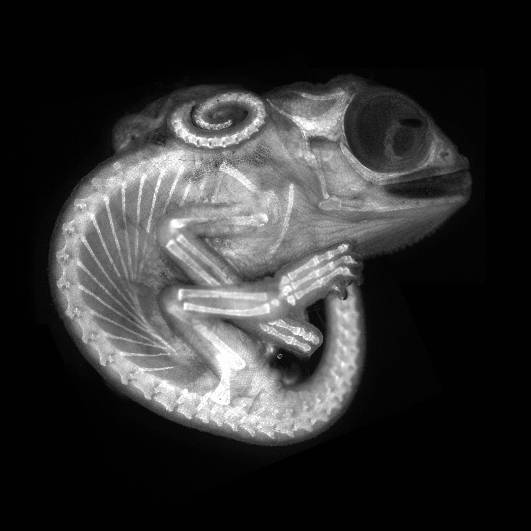
Chameleon embryo (autofluorescence). Dr. Allan Carrillo-Baltodano & David Salamanca (Queen Mary University of London, School of Biological and Chemical Sciences, London, United Kingdom). Eighth Place. Fluorescence, 10X (Objective Lens Magnification).


