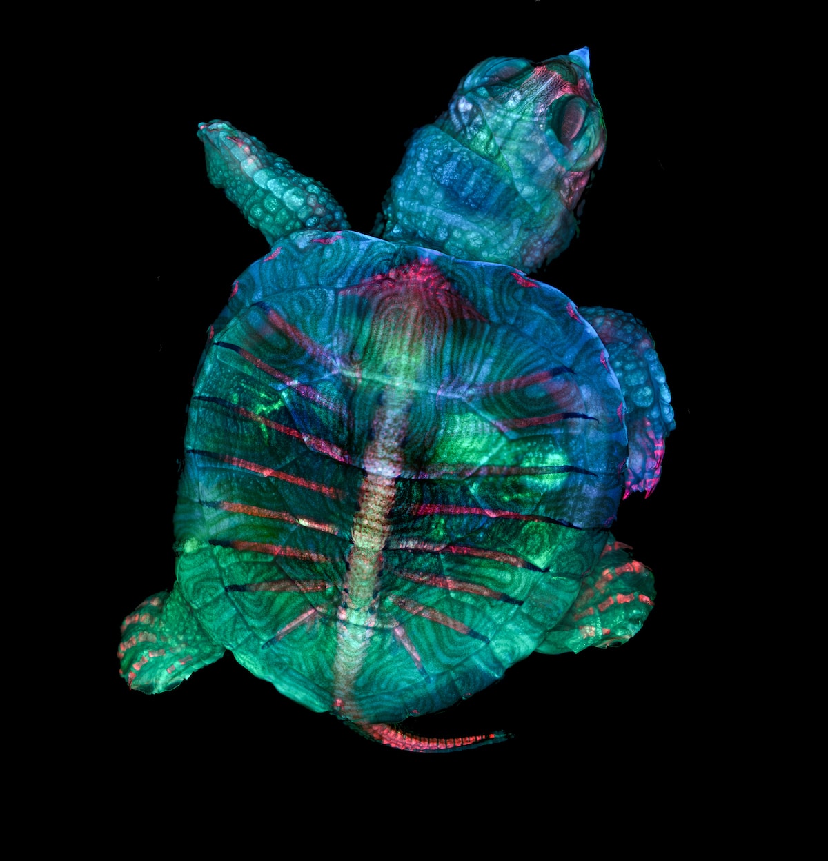
Fluorescent turtle embryo. Teresa Zgoda & Teresa Kugler (Campbell Hall, New York, USA). First Place. Stereomicroscopy, Fluorescence. 5x (Objective Lens Magnification).
For forty-five years, the Nikon Small World Photomicrography Competition has celebrated the microscopic world and, in the process, allowed scientists and enthusiasts to show off the artistry of scientific imagery. Passionate micro-photographers from nearly 100 countries submitted over 2000 stunning pieces of microphotography to the competition. In the end, the expert judging panel narrowed the field to the top 20 images with a photo of a turtle embryo taking the top prize.
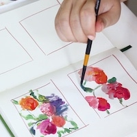
The colorful image, taken by microscopy technician Teresa Zgoda and recent university graduate Teresa Kugler, is the result of painstaking work done with precision and skill. Extensive image-stitching was necessary to create the final photograph, as the size and thickness of the turtle embryo meant that only a small portion of the turtle could be photographed at one time. By stacking and stitching together hundreds of photographs, the duo was able to create an image that is both scientifically and artistically satisfying.
“Microscopy lets us zoom in on the smallest organisms and building blocks that comprise our world–giving us a profound appreciation for the small things in life that far too often go unnoticed,” shared Kugler, “It allows me to do science with a purpose.”
The turtle wasn't the only embryo in the winning selection. Reproduction was a topic for many photographers. An alligator and a California two-spot octopus embryo, as well as a pregnant planktonic crustacean and mosquito larva also joined the top twenty. Away from the animal kingdom, something as simple as a frozen drop of water was transformed into a mesmerizing, abstract photograph. At the same time, a close view of different flora helps us marvel at the beauty and precision of how nature develops.
Whether using focus stacking, image stacking, or confocal microscopy, the techniques employed help these scientists bring their vision to life. And as technology continues to grow and evolve, we can only expect even richer results. Take a look at the rest of the top 20 photos from the 2019 Nikon Small World Photomicrography Competition below and view more finalists via their online gallery.
For 45 years, the Nikon Small World Photomicrography Competition has celebrated the artistry of scientific imagery.
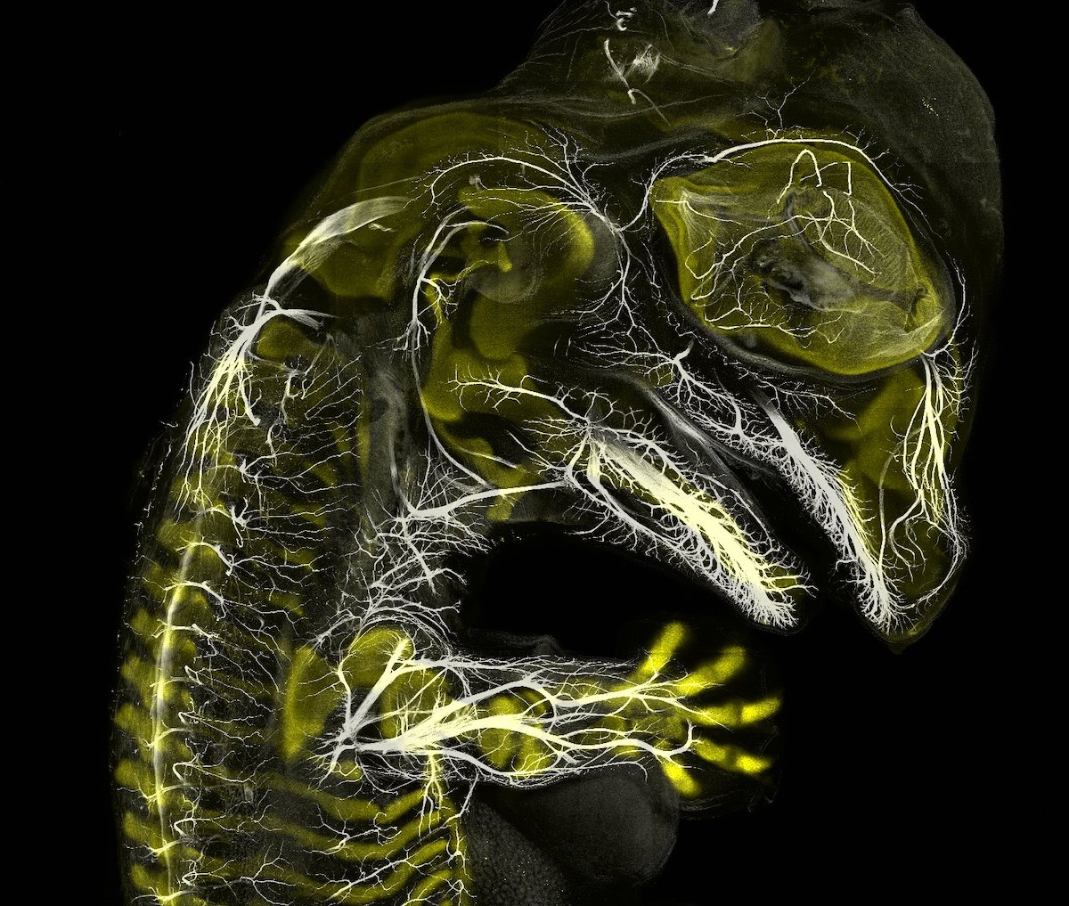
Alligator embryo developing nerves and skeleton. Daniel Smith Paredes & Dr. Bhart-Anjan S. Bhullar (Yale University, Department of Geology and Geophysics, New Haven, Connecticut, USA). Third Place. Immunofluorescence. 10x (Objective Lens Magnification).
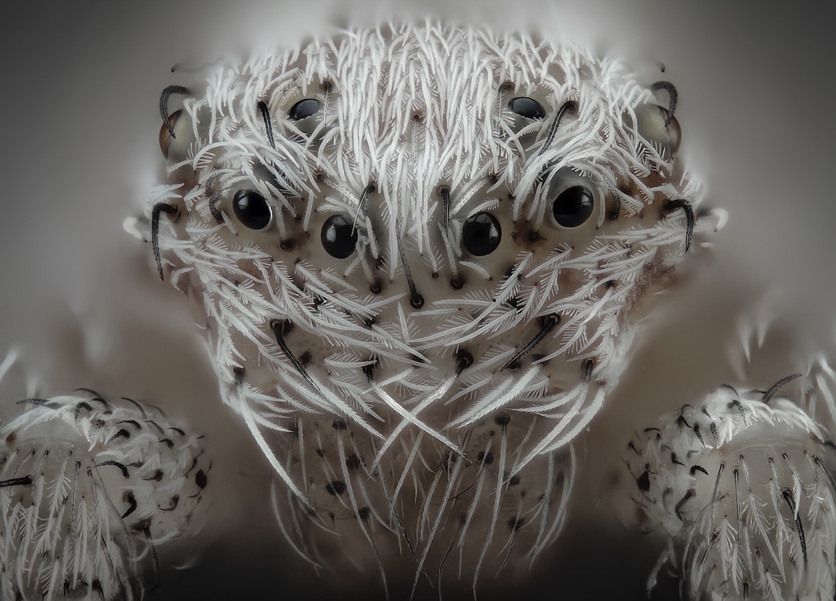
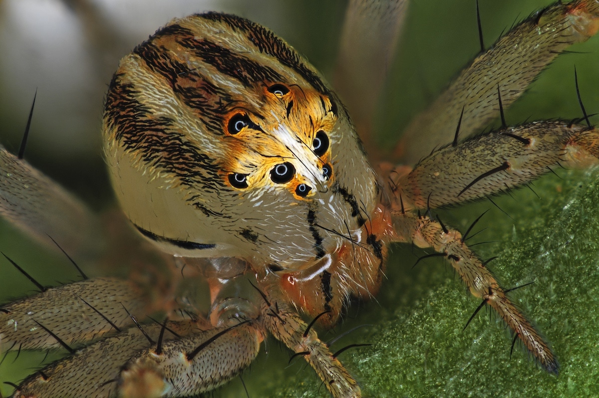
Small white hair spider. Javier Rupérez (Almáchar, Málaga, Spain). Sixth Place. Reflected Light, Image Stacking. 20x (Objective Lens Magnification).
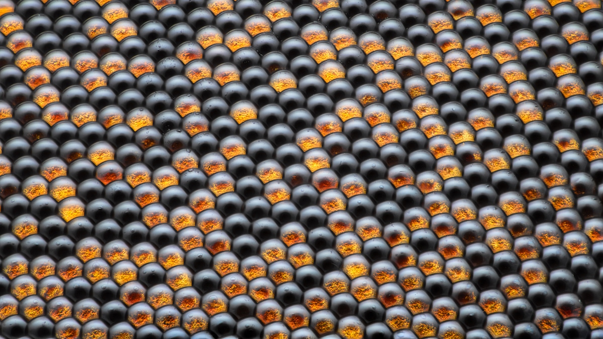
Housefly compound eye pattern. Dr. Razvan Cornel Constantin (Bucharest, Romania). 16th Place. Focus Stacking, Reflected Light. 50x (Objective Lens Magnification).
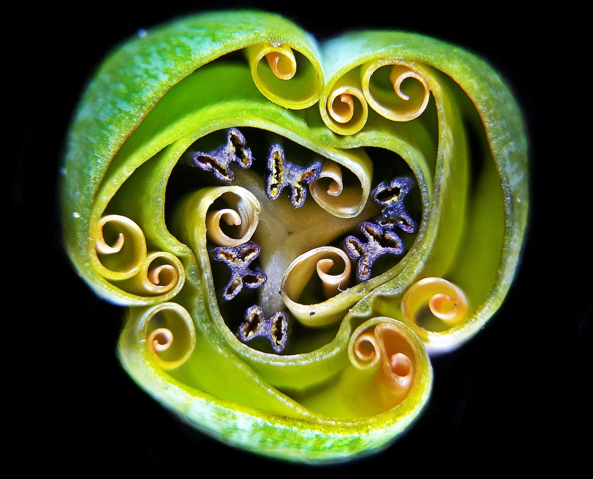
Tulip bud cross section. Andrei Savitsky (Cherkassy, Ukraine). Ninth Place. Reflected Light. 1x (Objective Lens Magnification).
Female Oxyopes dumonti (lynx) spider. Antoine Franck (CIRAD – Agricultural Research for Development, Saint Pierre, Réunion). 14th Place. Focus Stacking. 1x (Objective Lens Magnification).
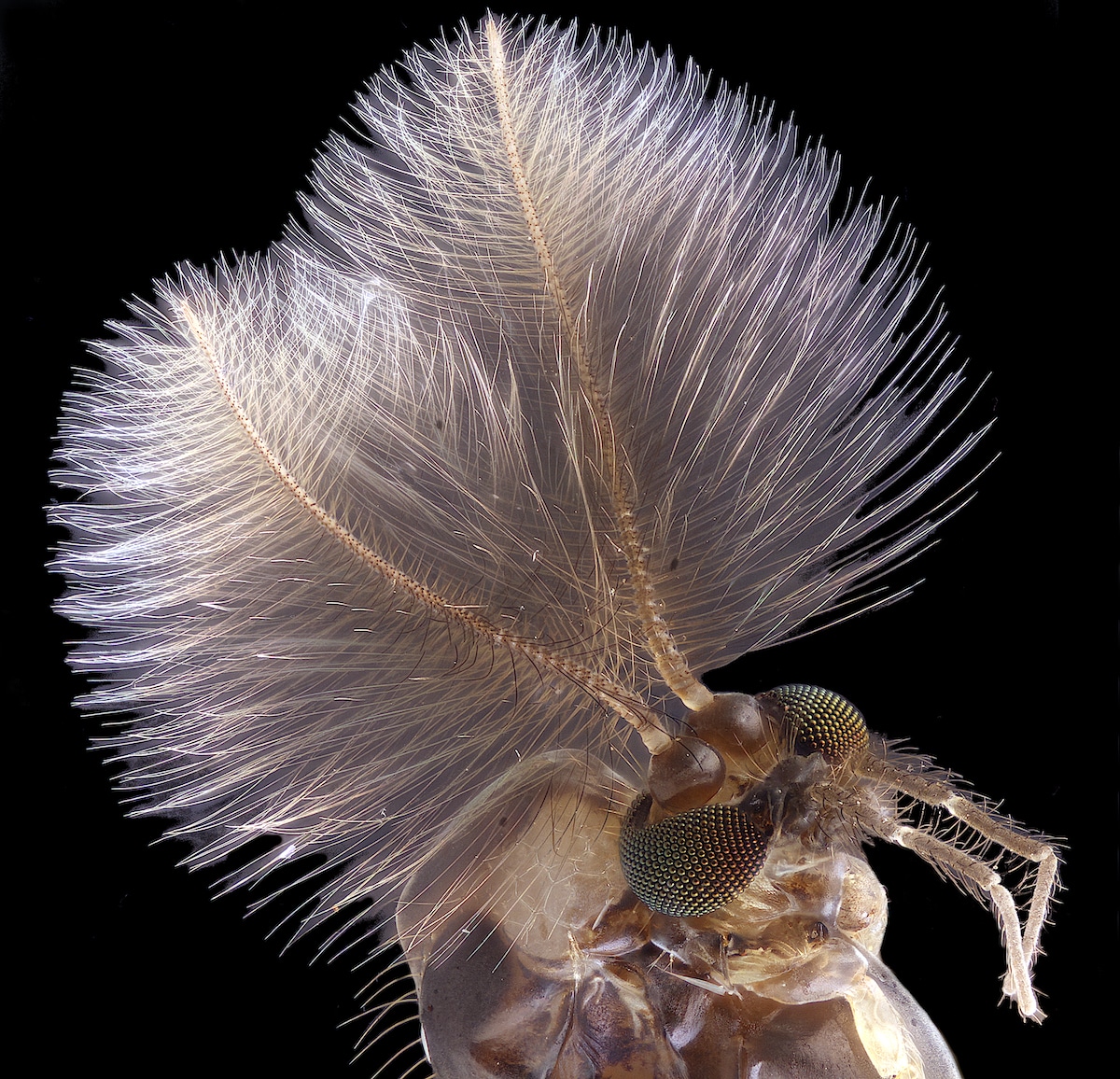
Male mosquito. Jan Rosenboom (Universität Rostock, Rostock, Mecklenburg Vorpommern, Germany). Fourth Place. Focus Stacking. 6.3x (Objective Lens Magnification).
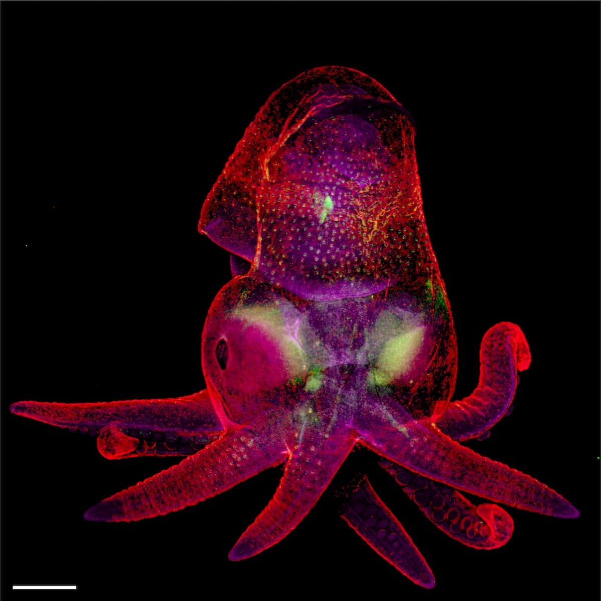
Octopus bimaculoides embryo. Martyna Lukoseviciute & Dr. Carrie Albertin (University of Oxford, Weatherall Institute of Molecular Medicine, Oxford, Oxfordshire, United Kingdom). 19th Place. Confocal, Image Stitching. 5x (Objective Lens Magnification).
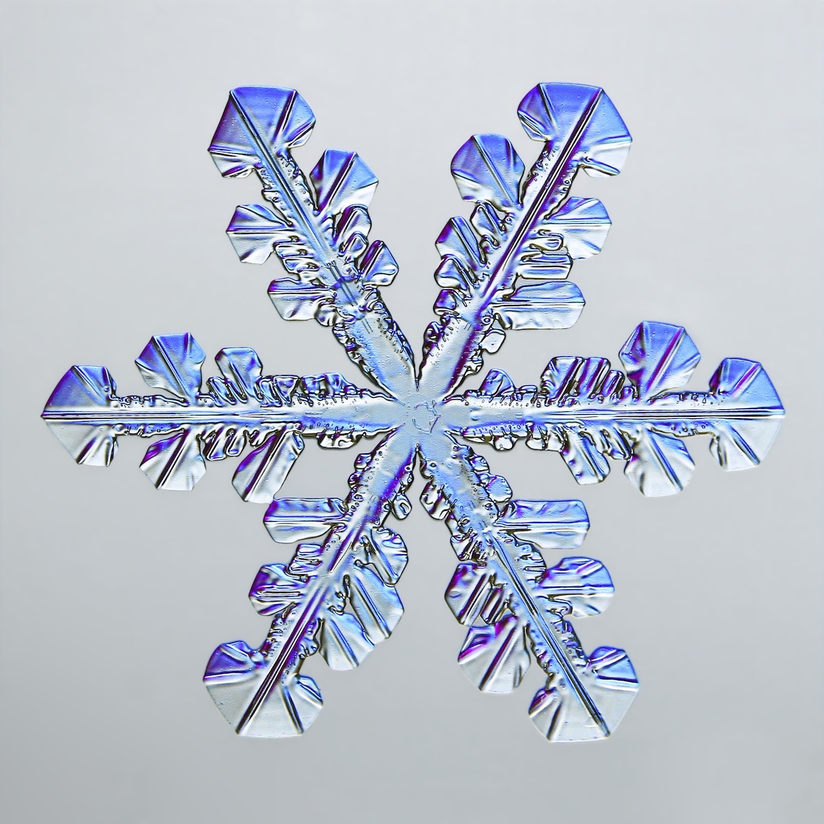
Snowflake. Caleb Foster (Caleb Foster Photography, Jericho, Vermont, USA). Fifth Place. Transmitted Light. 4x (Objective Lens Magnification).
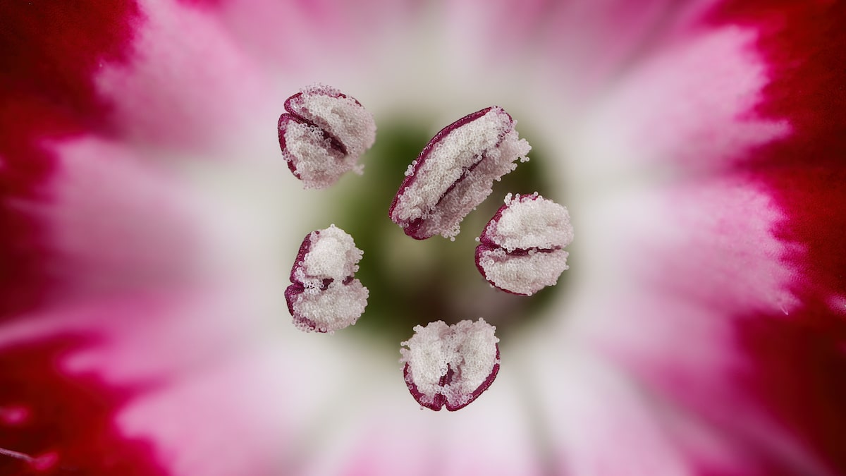
Chinese red carnation stamen. Dr. Guillermo López López (Alicante, Spain). Seventh Place. Focus Stacking. 3x (Objective Lens Magnification).
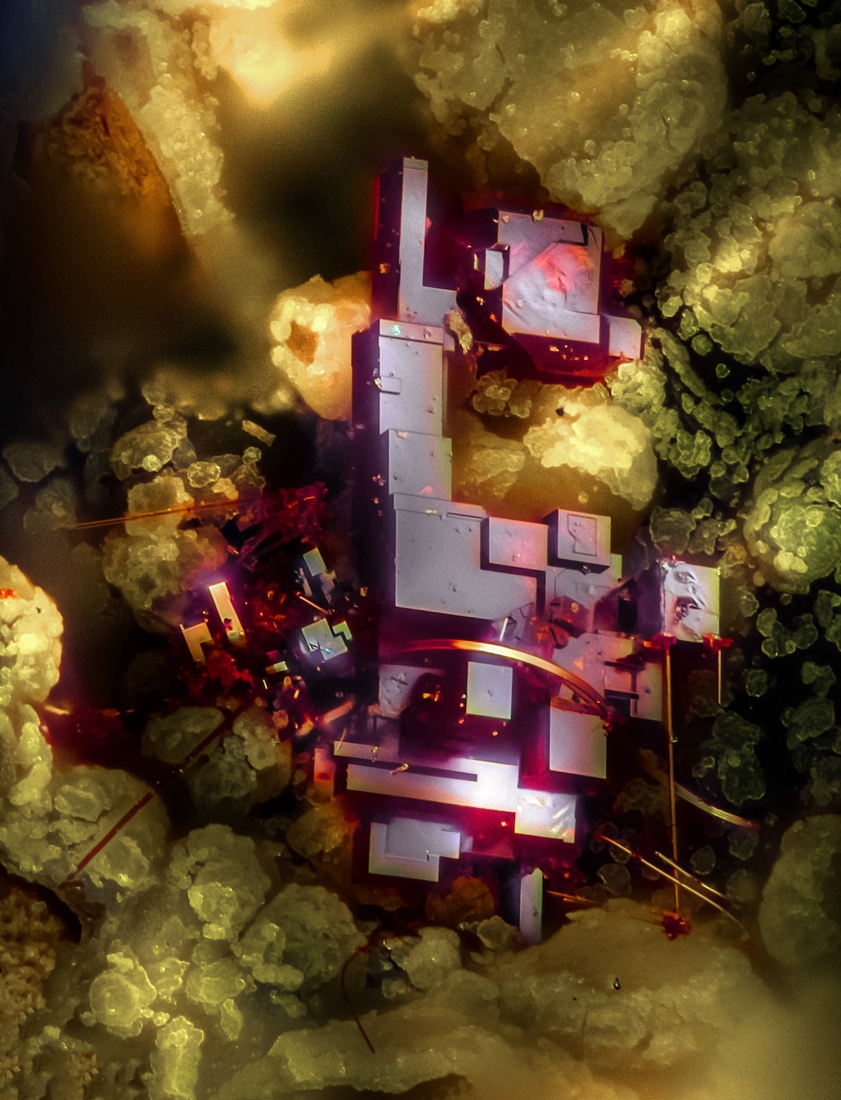
Cuprite (mineral composed of copper oxide). Dr. Emilio Carabajal Márquez (Madrid, Spain). Focus Stacking. 20x (Objective Lens Magnification).
Vitamin C. Karl Deckart (Eckental, Bavaria, Germany). 17th Place. Brightfield, Polarized Light. 4x (Objective Lens Magnification)
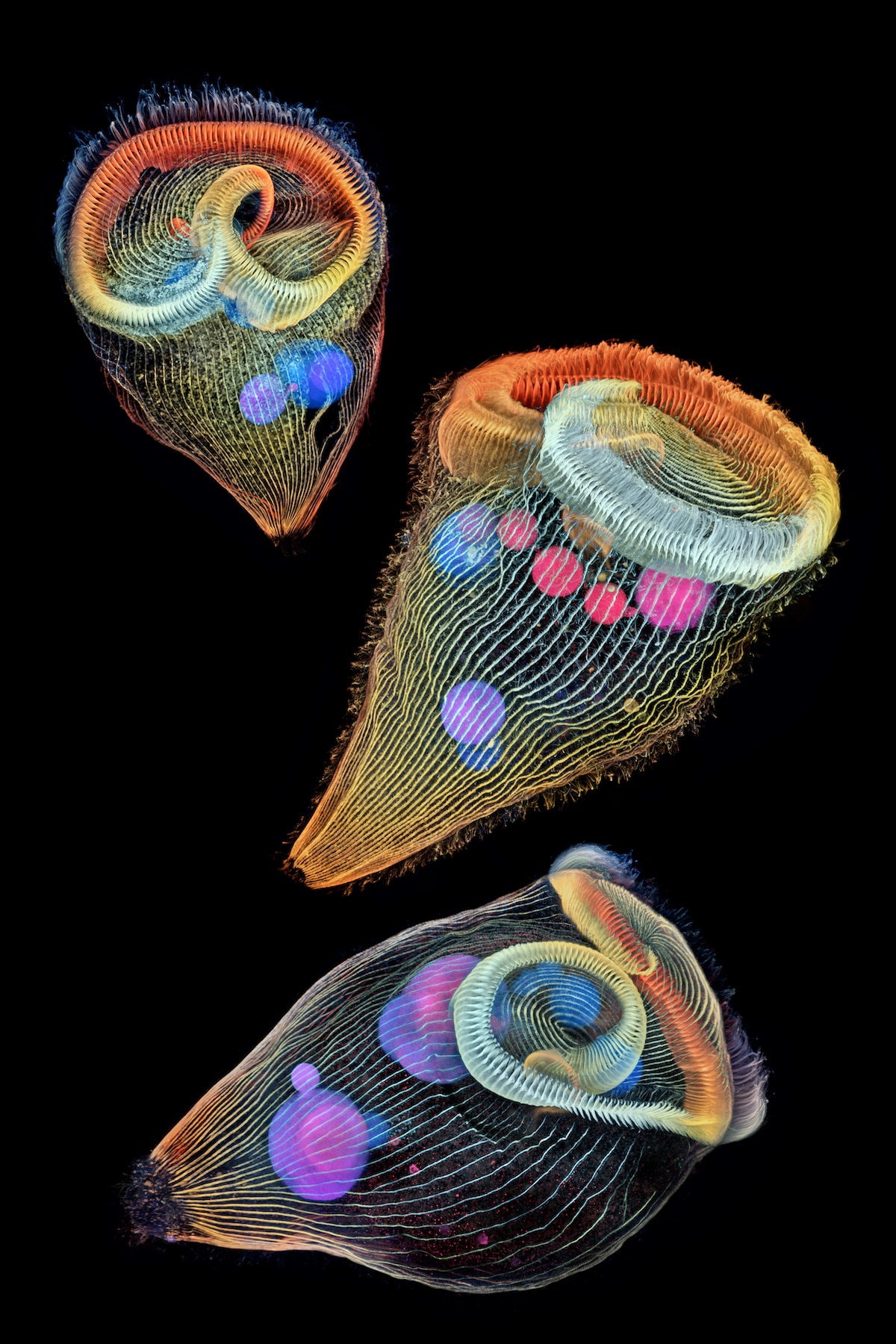
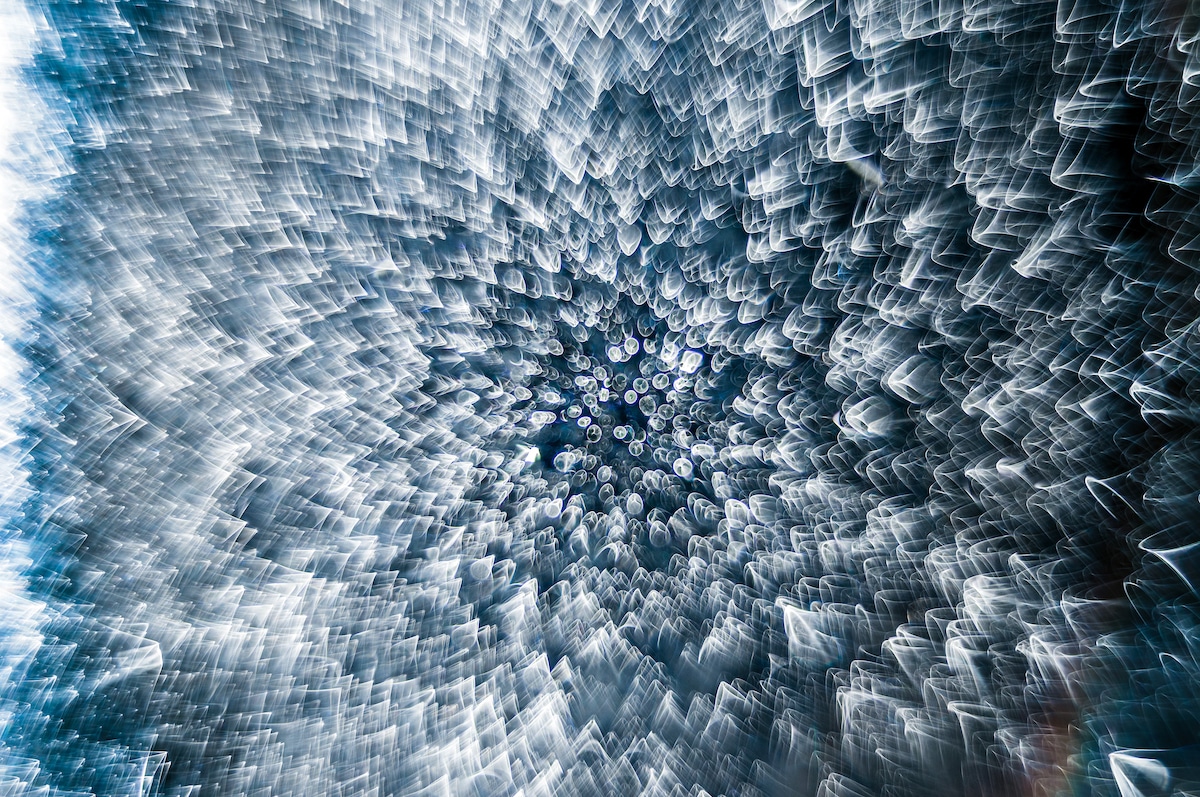
Depth-color coded projections of three stentors (single-cell freshwater protozoans). Dr. Igor Siwanowicz (Howard Hughes Medical Institute (HHMI), Janelia Research Campus, Ashburn, Virginia, USA). Second place. Confocal. 40x (Objective Lens Magnification).
Frozen water droplet. Garzon Christian (Quintin, Cotes-d’Armor, France). Eighth Place. Incident Light. 8x (Objective Lens Magnification).
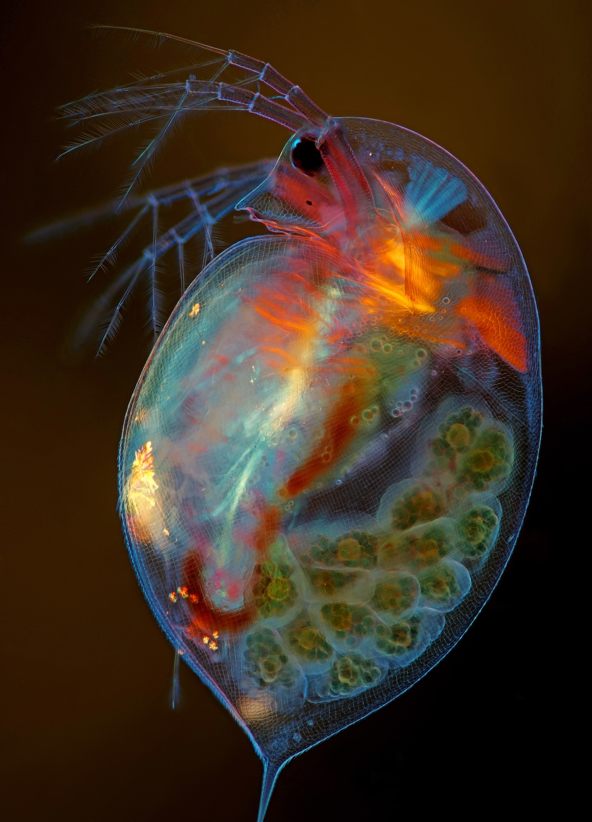
Pregnant Daphnia magna (small planktonic crustacean). Marek Miś (Marek Miś Photography, Suwalki, Podlaskie, Poland). 15th Place. Modified Darkfield, Polarized Light, Image Stacking. 4x (Objective Lens Magnification).
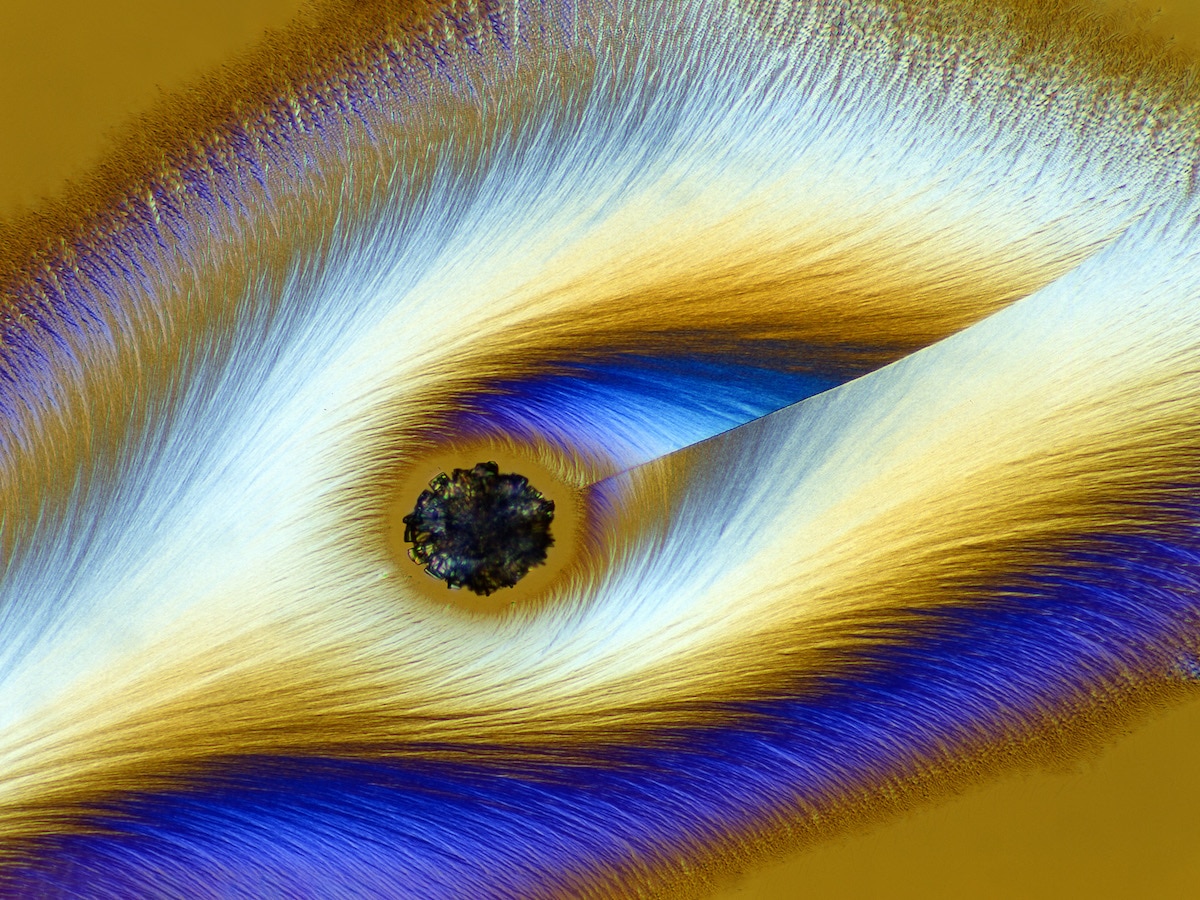
Cristobalite crystal suspended in its quartz mineral host. E. Billie Hughes (Lotus Gemology, Bangkok, Thailand). 18th Place. Darkfield. 40x (Objective Lens Magnification).
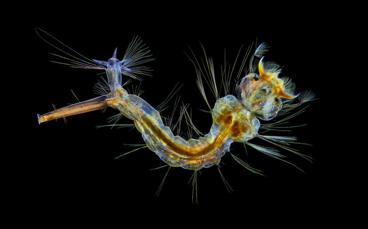
Mosquito larva. Anne Algar (Hounslow, Middlesex, United Kingdom). 12th Place. Darkfield, Polarizing Light, Image Stacking. 4x (Objective Lens Magnification).
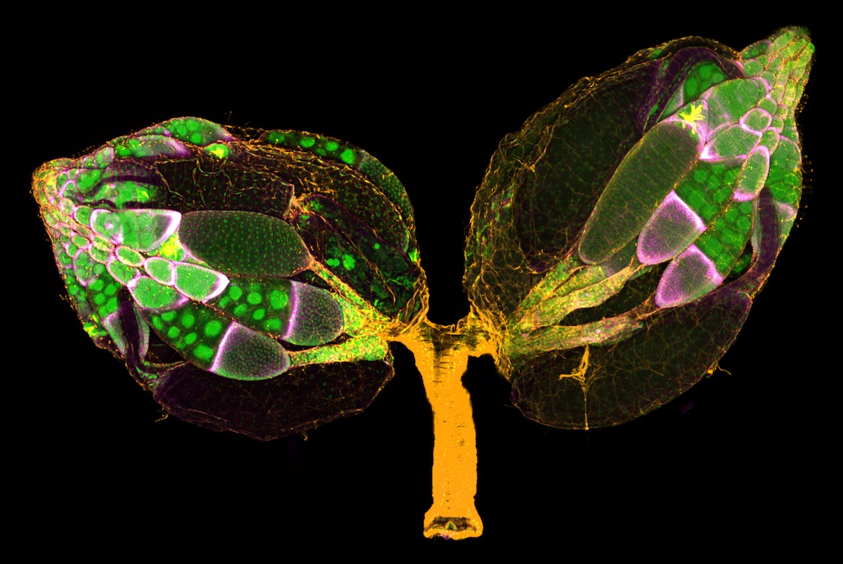
A pair of ovaries from an adult Drosophila female stained for F-actin (yellow) and nuclei (green); follicle cells are marked by GFP (magenta). Dr. Yujun Chen & Dr. Jocelyn McDonald (Kansas State University, Department of Biology, Manhattan, Kansas, USA). 11th Place. Confocal. 10x (Objective Lens Magnification).
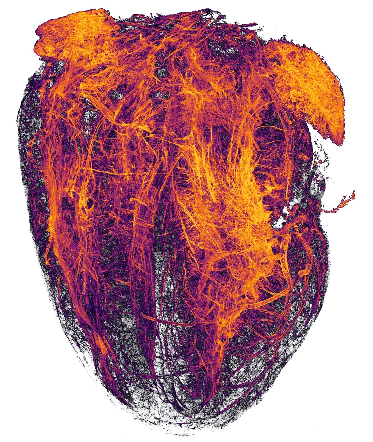
Blood vessels of a murine (mouse) heart following myocardial infarction (heart attack). Simon Merz, Lea Bornemann & Sebastian Korste (University Hospital Essen, Institute for Experimental Immunology & Imaging, Essen, Nordrhein-Westfalen, Germany). 20th Place. Tissue Clearing, Light Sheet Fluorescence Microscopy. 2x (Objective Lens Magnification).
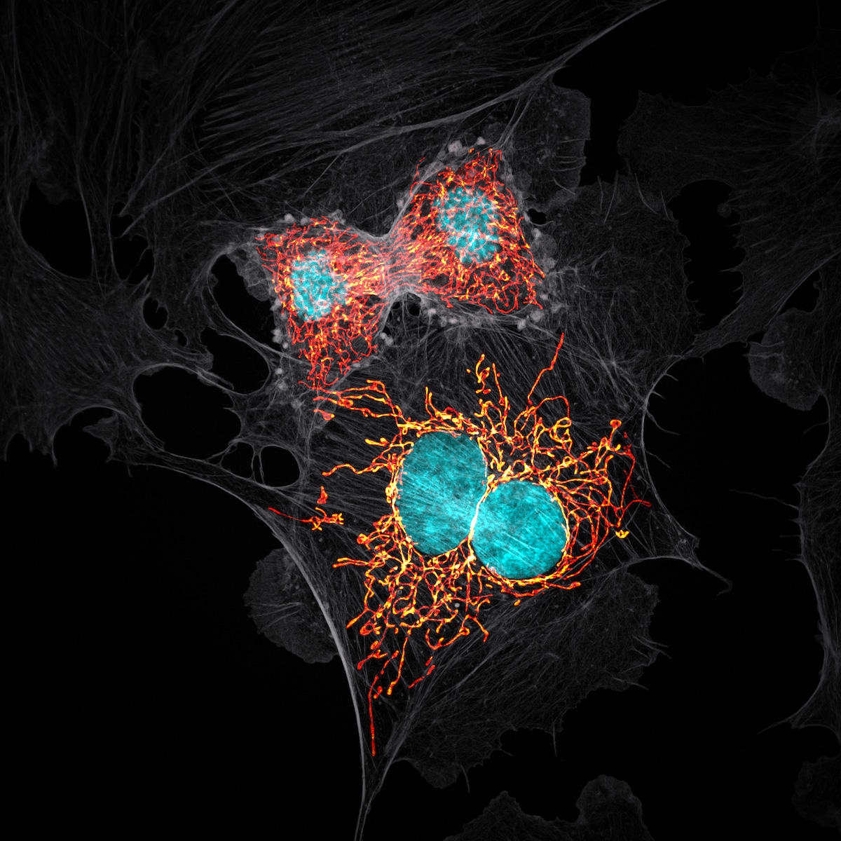
BPAE cells in telophase stage of mitosis. Jason M. Kirk (Baylor College of Medicine, Optical Imaging & Vital Microscopy Core, Houston, Texas, USA). 10th Place. Confocal with Enhanced Resolution. 63x (Objective Lens Magnification).