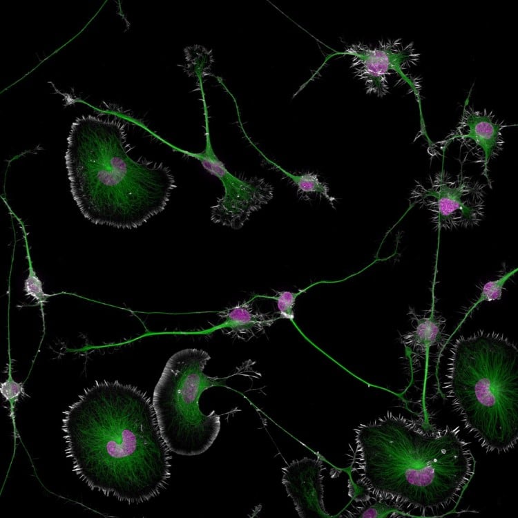
1st Place, Dr. Bruno Cisterna & Dr. Eric Vitriol, Medical College of Georgia at Augusta University, Department of Neuroscience & Regenerative Medicine. Differentiated mouse brain tumor cells (actin, microtubules, and nuclei), Super-Resolution, 40X (Objective Lens Magnification)
Known for recognizing excellence in photomicrography—photography through a microscope—the Nikon Small World competition is celebrating its 50th anniversary. For five decades, this unique photo contest has highlighted the beauty of the microscopic world, bringing it to a wider public and evolving with advancements in technology.
This year, 2,100 photos from 80 countries were entered into the competition and judged on originality, informational content, technical proficiency, and visual impact. Dr. Bruno Cisterna and Dr. Eric Vitriol of Augusta University's Medical College of Georgia were named the winners for their fascinating look at a differentiated mouse brain tumor. Their image demonstrates how disruptions in the cell's cytoskeleton—the structural framework and “highways” known as microtubules—can lead to diseases like Alzheimer's and ALS.
“One of the main problems with neurodegenerative diseases is that we don't fully understand what causes them,” shares Dr. Cisterna. “To develop effective treatments, we need to figure out the basics first. Our research is crucial for uncovering this knowledge and ultimately finding a cure. Differentiated cells could be used to study how mutations or toxic proteins that cause Alzheimer's or ALS alter neuronal morphology, as well as to screen potential drugs or gene therapies aimed at protecting neurons or restoring their function.”
The second and third-place winners exemplify the diversity found in the competition. Dr. Marcel Clemens took home second place for his striking image of an electrical arc between a pin and a wire, produced by applying a potential difference of 10,000 volts. Third place was handed out to Chris Romaine for his fascinating look at a cannabis leaf with bulbous trichomes.
“At 50 years, Nikon Small World is more than just an imaging competition—it’s become a gallery that pays tribute to the extraordinary individuals who make it possible. They are the driving force behind this event, masterfully blending science and art to reveal the wonders of the microscopic world and what we can learn from it to the public,” shares Eric Flem, Senior Manager, CRM and Communications at Nikon Instruments.
“Sometimes, we overlook the tiny details of the world around us. Nikon Small World serves as a reminder to pause, appreciate the power and beauty of the little things, and to cultivate a deeper curiosity to explore and question.”
Scroll down to see the top 20 images and then head over to the official website to see all 87 recognized photos from the 2024 competition.
Here are the winners of the 2024 Nikon Small World Photomicrography Competition.

2nd Place, Dr. Marcel Clemens, Verona, Veneto, Italy. Electrical arc between a pin and a wire
Image stacking for the pin and wire combined with long exposure for the electrical arcs, 10X (Objective Lens Magnification)
3rd Place, Chris Romaine, Kandid Kush, Port Townsend, Washington, USA. Leaf of a cannabis plant. The bulbous glands are trichomes. The bubbles inside are cannabinoid vesicles, Image Stacking, 20X (Objective Lens Magnification)
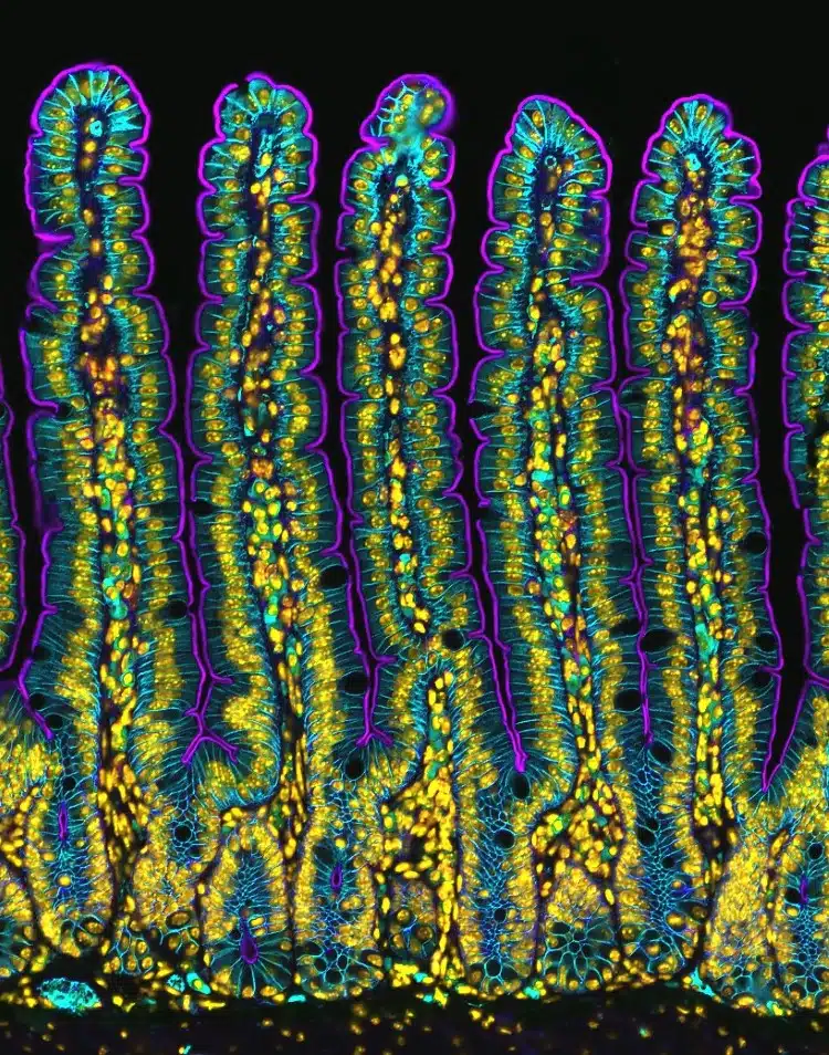
4th Place, Dr. Amy Engevik, Medical University of South Carolina Department of Regenerative Medicine & Cell Biology. Section of a small intestine of a mouse, Fluorescence
10X (Objective Lens Magnification)
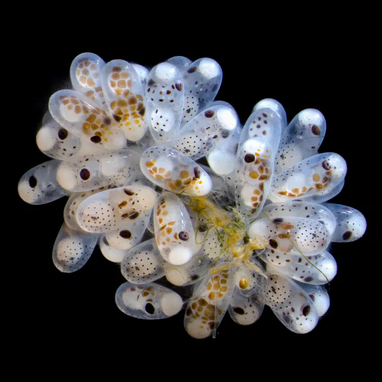
5th Place, Thomas Barlow & Connor Gibbons, Columbia University Department of Neurobiology and Behavior. Cluster of octopus (Octopus hummelincki) eggs, Darkfield, Stereomicroscopy, Focus Stacking, 3X (Objective Lens Magnification)
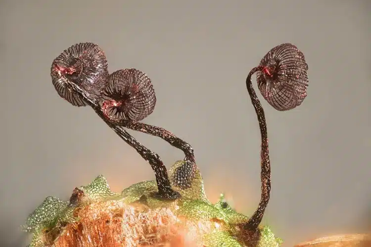
6th Place, Henri Koskinen, Helsinki University. Slime mold (Cribraria cancellata)
Image Stacking, Polarized Light, Reflected Light, 10X (Objective Lens Magnification)
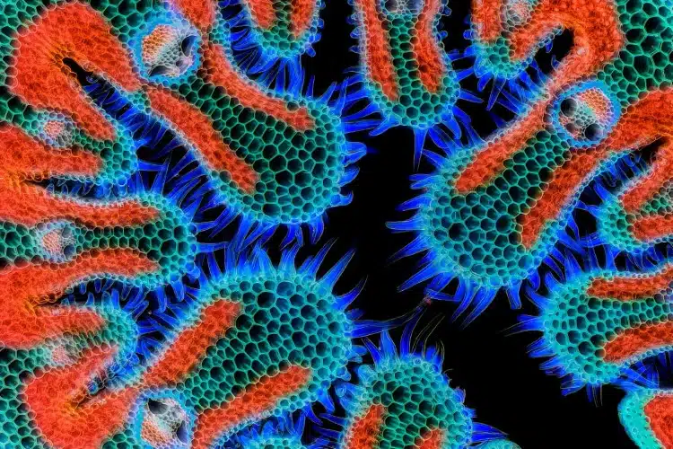
7th Place, Gerhard Vlcek, Maria Enzersdorf, Austria. Cross section of European beach grass (Ammophila arenaria) leaf, Brightfield, Image Stacking, 10X (Objective Lens Magnification)
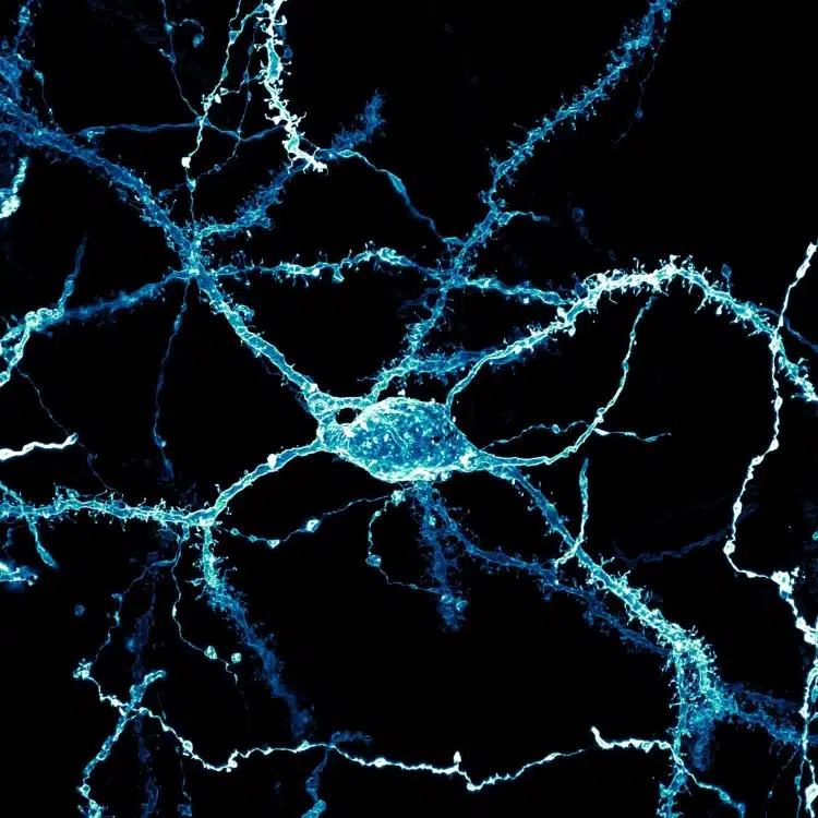
8th Place, Stephanie Huang, Victoria University of Wellington, School of Biological Sciences; School of Psychology
Wellington, New Zealand. A neuron densely covered in dendritic spines from the striatum of an adult rat brain, Confocal, Deconvolution, Image Stacking, 60X (Objective Lens Magnification)
The prestigious photo contest recognizes excellence in photography through the microscope.
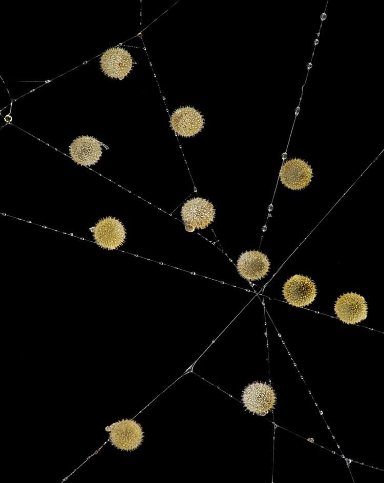
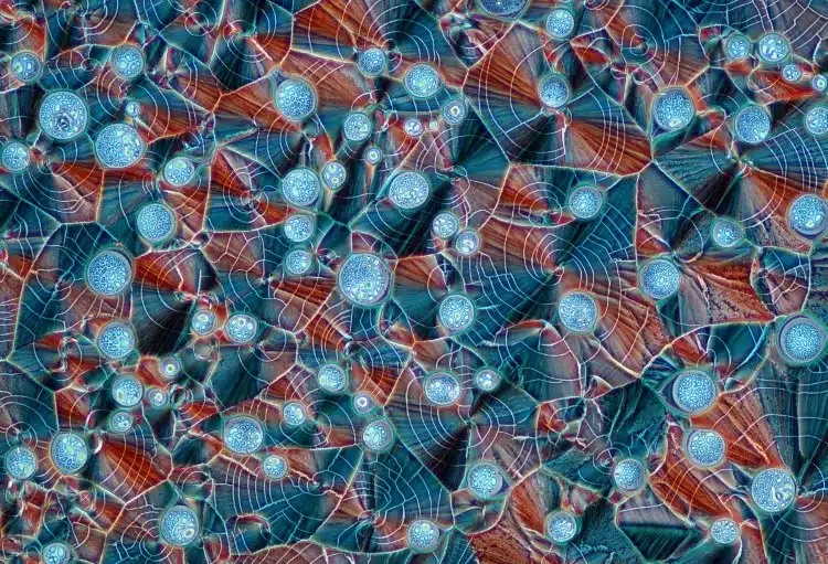
9th Place, John-Oliver Dum, Medienbunker Produktion, Bendorf, Rheinland Pfalz, Germany. Pollen in a garden spider (Araneus) web, Image Stacking, 20X (Objective Lens Magnification)
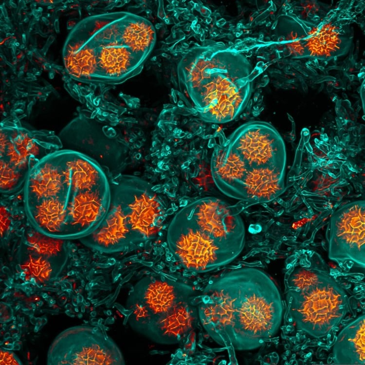
10th Place, Jan Martinek, Charles University Department of Experimental Plant Biology, Prague, Czech Republic. Spores of black truffle (Tuber melanosporum), Confocal, 63X (Objective Lens Magnification)
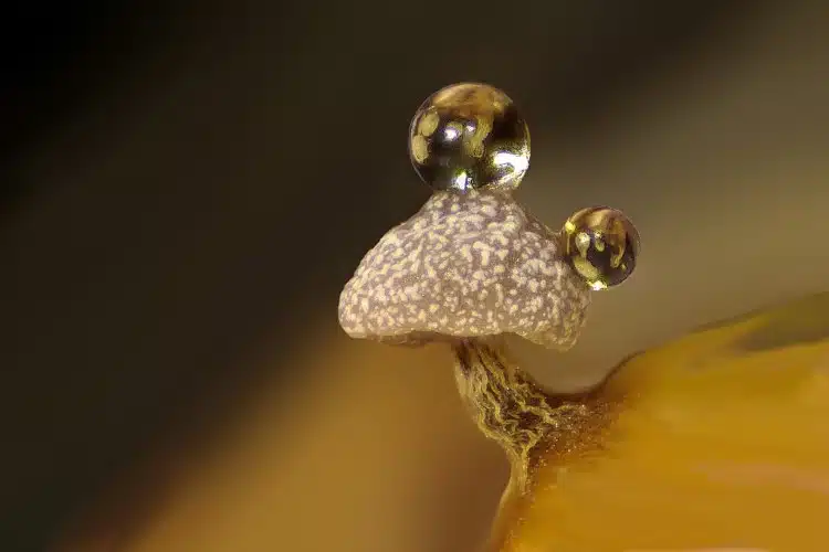
11th Place, Dr. Ferenc Halmos, Bánd, Veszprém, Hungary. Slime mold on a rotten twig with water droplets, Image Stacking, 0.7X – 4.5X (Objective Lens Magnification)
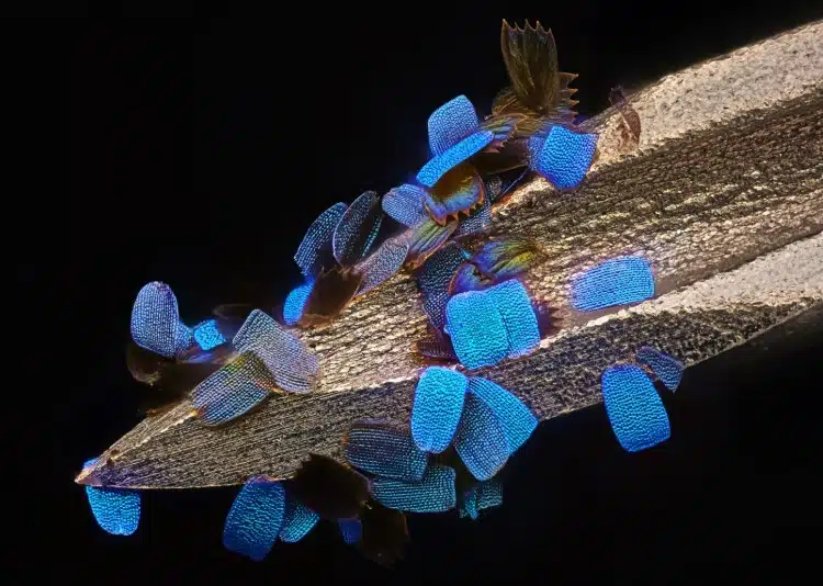
12th Place, Daniel Knop, Oberzent-Airlenbach, Hessen, Germany. Wing scales of a butterfly (Papilio ulysses) on a medical syringe needle, Image Stacking, 20X (Objective Lens Magnification)
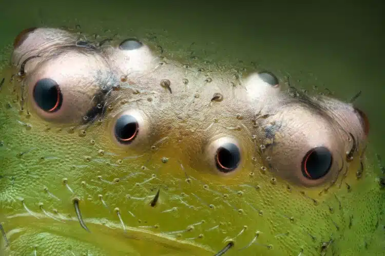
13th Place, Paweł Błachowicz, Bedlno, Świętokrzyskie, Poland. Eyes of green crab spider (Diaea dorsata), Image Stacking, Reflected Light, 20X (Objective Lens Magnification)
14th Place, Marek Miś, Marek Miś Photography, Suwalki, Podlaskie, Poland. Recrystallized mixture of hydroquinone and myoinositol, Polarized Light, 10X (Objective Lens Magnification)
This year the contest celebrates its 50th anniversary.
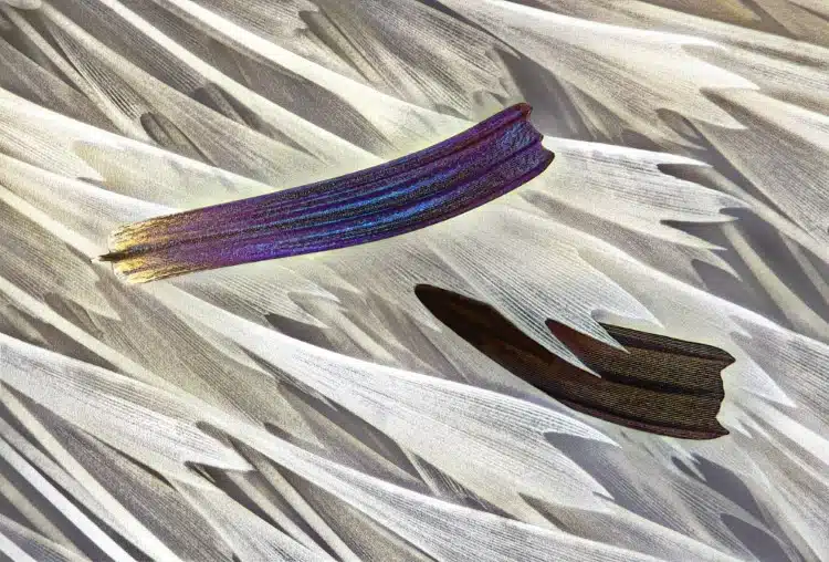
15th Place, Sébastien Malo, Saint Lys, Haute-Garonne, France. Isolated scales on Madagascan sunset moth wing (Chrysiridia ripheus), Darkfield, Image Stacking, Reflected Light, 40X (Objective Lens Magnification)
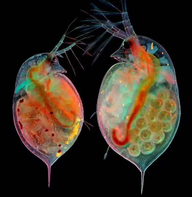
16th Place, Marek Miś, Marek Miś Photography, Suwalki, Podlaskie, Poland. Two water fleas (Daphnia sp.) with embryos (left) and eggs (right), Darkfield, Polarized Light, 10X (Objective Lens Magnification)
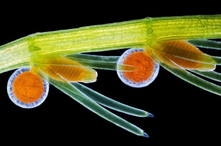
17th Place, Dr. Frantisek Bednar, Svosov, Zilinsky, Slovak Republic. Stonewort algae (Chara virgata) reproductive organs – oogonia (female organs) and antheridia (male organs), Darkfield, 4X (Objective Lens Magnification)
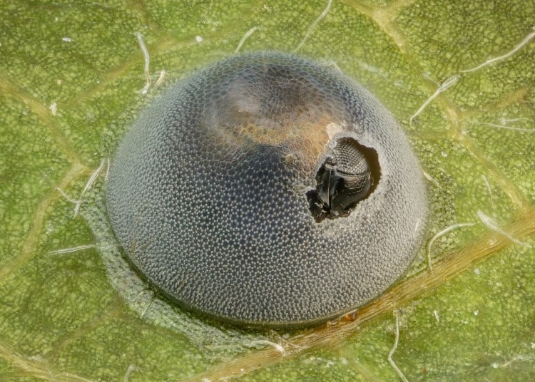
18th Place, Alison Pollack, San Anselmo, California, USA. An insect egg parasitized by a wasp, Image Stacking, Reflected Light, 10X (Objective Lens Magnification)
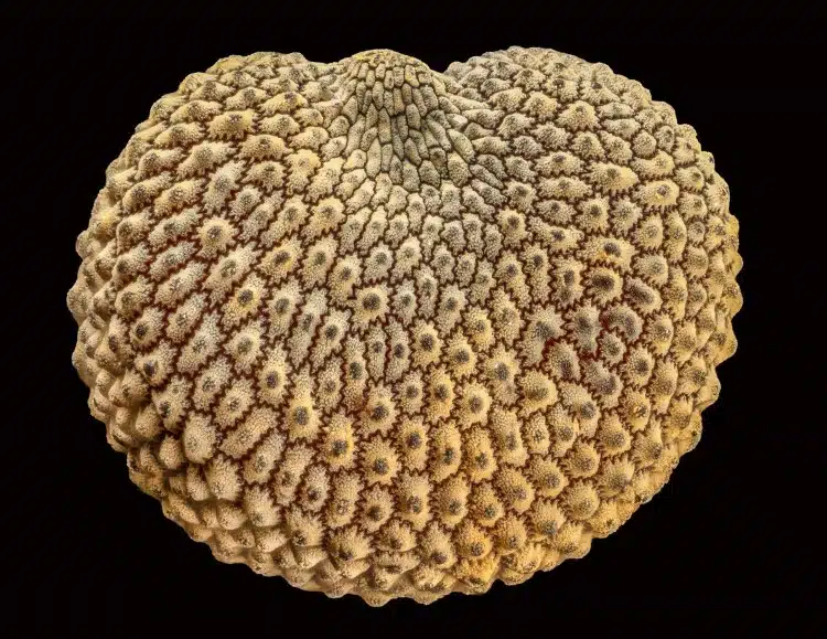
19th Place, Alison Pollack, San Anselmo, California, USA. Seed of a Silene plant, Image Stacking, Reflected Light, 10X (Objective Lens Magnification)
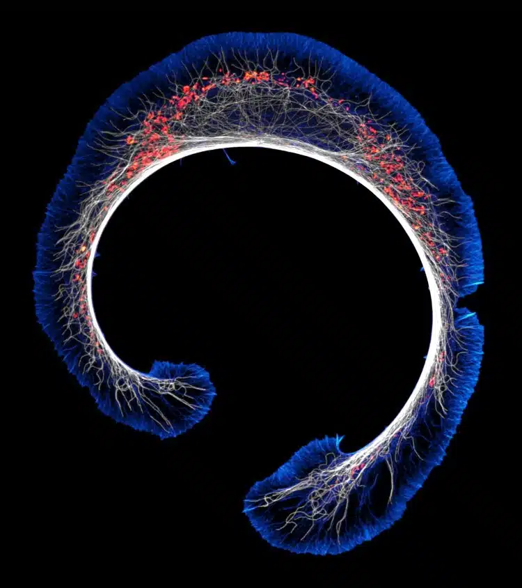
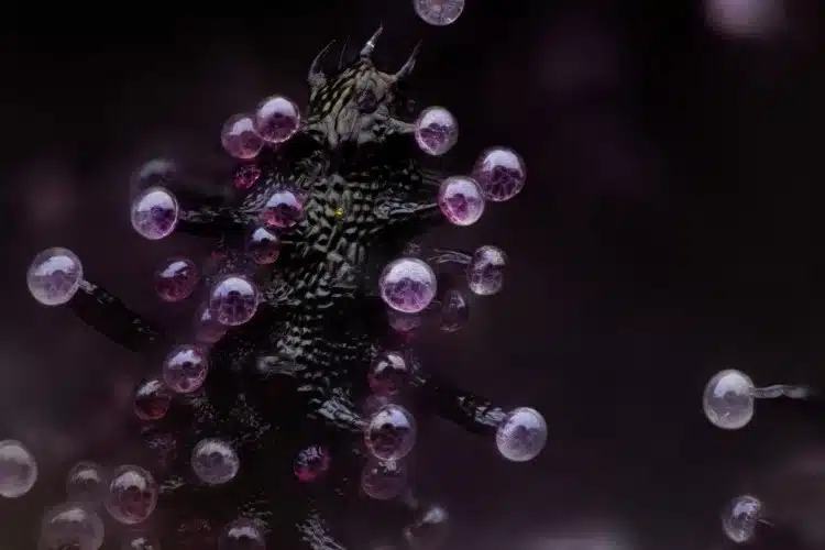
20th Place, Dr. Bruno Cisterna & Dr. Eric Vitriol, Medical College of Georgia at Augusta University
Department of Neuroscience & Regenerative Medicine. Early stage of mouse glioblastoma cell differentiation (actin, microtubules, and mitochondria), Super-Resolution, 100X (Objective Lens Magnification)
Nikon Small World: Website | Facebook | Instagram
My Modern Met granted permission to feature photos by Nikon Small World.
Related Articles:
Glowing Frog on a Bioluminescent Mushroom Wins Science Photography Contest
Psychedelic Image of Optic Nerve Wins Nikon Small World Photomicrography Contest
Winners of the Nikon Small World Photomicrography Contest Create Art Using a Microscope
Explore Life Under the Microscope With the Winners of the Nikon Small World Photomicrography Contest